Safe zones for pin placement in the pediatric femur
1. Introduction
The safest anatomical zones for pin insertion are the anterolateral and direct lateral regions of the femur.
2. General considerations
Anatomy
The thigh is covered by a circumferential muscular envelope and the diaphysis of the femur by a thick periosteum.
The major neurovascular structures are located medially and, therefore, the femur can be safely approached over the anterolateral region.
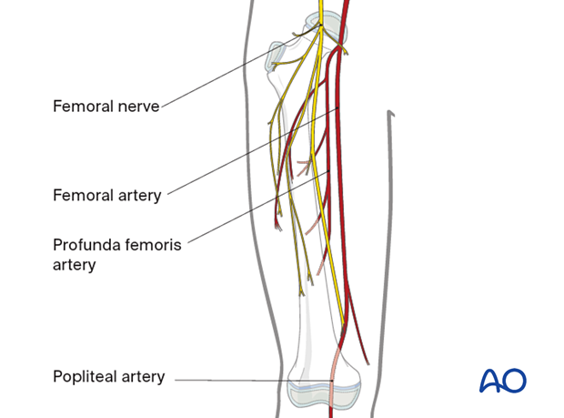
Overall neurovascular status of the limb
For the pin insertion, consider the state of the soft-tissue envelope of the femoral shaft (areas of skin loss or extensive soft-tissue damage should be avoided, to minimize the risk of subsequent pin-track infection).
Conversion of temporary external fixator to nailing or plating
Take care to protect the skin and soft tissues during pin insertion, whether the external fixator is to be used as a definitive device or substituted for an intramedullary nail or plate at a later stage.
3. Safe zone in the proximal third
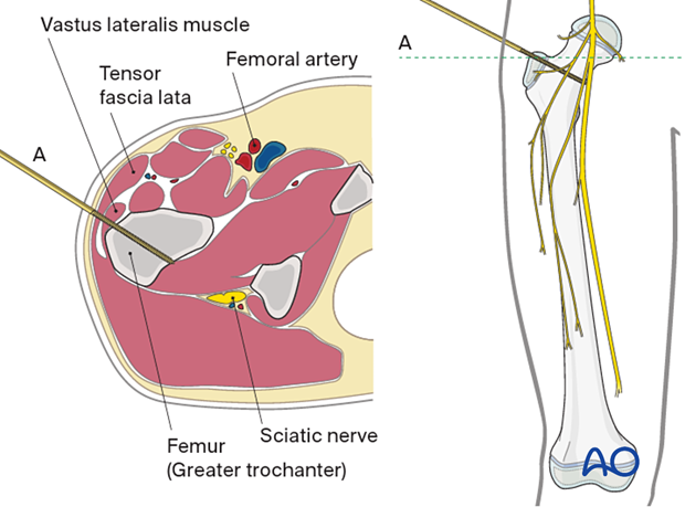
With the patient supine, palpate the greater trochanter and, depending on the fracture configuration, direct the pin through vastus lateralis, aiming towards the lesser trochanter (A) …
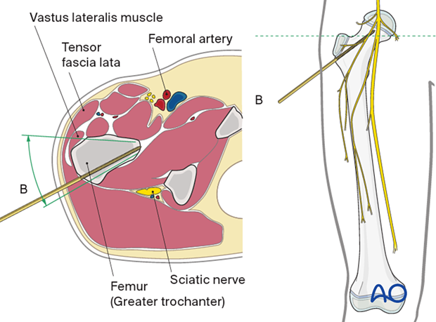
… or the femoral neck (B).
Ensure that the entry points for pin insertion will not subsequently conflict with the conversion of the external fixator to an intramedullary nail.
The safe zone for pin placement includes the area from just behind the anterior margin to the posterior margin of the lateral face of the greater trochanter.
4. Safe zone in the midshaft
The anatomy in the area between the two solid lines is represented by one cross-section and does not vary significantly at different levels within this zone.
The direct lateral approach is preferred because it interferes the least with knee motion.
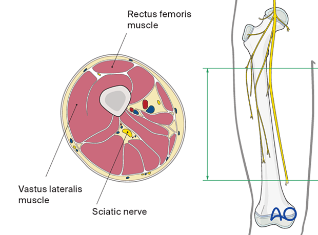
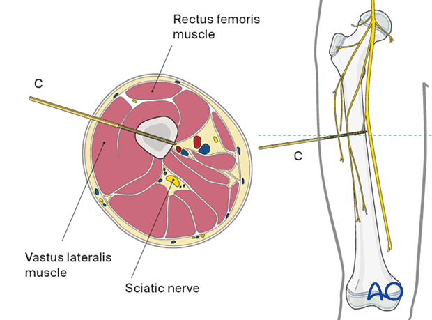
Direct lateral approach (C)
Palpate the vastus lateralis and insert the pins in the direction shown in the diagram, aiming to obtain purchase in both cortices.
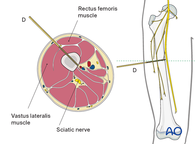
Anterolateral approach (D)
Palpate vastus lateralis and rectus femoris with the patient in supine position. The direction of the pin should be in the plane between these two muscles, as shown in the cross-section illustration. Take care to avoid perforation beyond the femoral cortex medially to prevent injury to the neurovascular structures.
5. Safe zone in the distal third
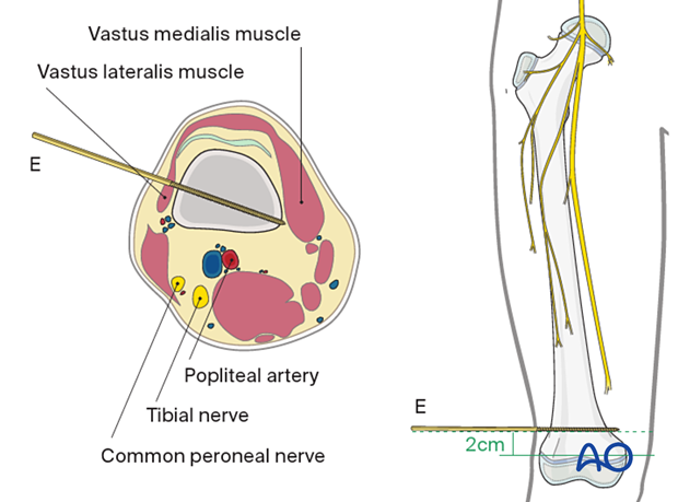
Direct lateral approach (E)
The lateral area of the distal femur is easily accessible for pin insertion. The inferior border of vastus lateralis is the only soft-tissue structure to avoid.
The pin should be positioned at least 2 cm proximal to the growth plate.


