Entry point in the pediatric femur for lateral-entry intramedullary nailing
1. Introduction
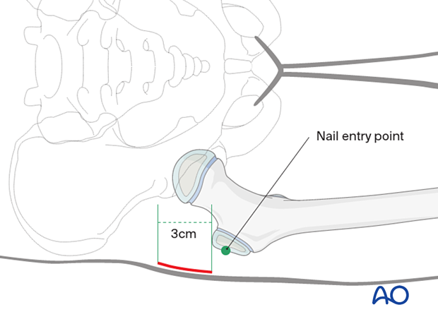
The nail entry point lies on the lateral surface of the greater trochanter.
2. Skin incision
The skin incision is centered on the palpable tip of the greater trochanter and extends 3 cm proximally, and distally as required.

The incision should be in line with the medullary canal on the lateral view, which can be marked on the skin using the lateral image intensifier view.

3. Deep dissection
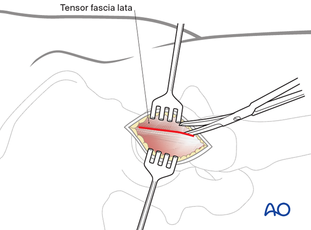
Incise the fascia lata in the same direction to expose the underlying greater trochanter.

4. Determination of entry point
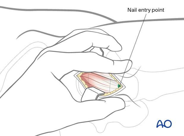
The entry point of the nail lies lateral and inferior to the tip of the greater trochanter. This can be clearly seen on an AP image-intensifier view.
Carefully palpate the anterior and posterior borders of the greater trochanter to identify its center.

5. Wound closure
Close the wound in a standard manner.


