Traction with delayed spica casting
1. General considerations
Introduction
Treatment with traction requires skillful application of the device and careful attention to detail with constant monitoring throughout the treatment period to avoid skin problems or vascular compromise.
Once the child is comfortable and moving or callus formation is visible on x-ray remove the traction and convert into a spica cast.
Traction types
There are a variety of devices that depend on the patient’s age, weight, fracture configuration, and resources available.
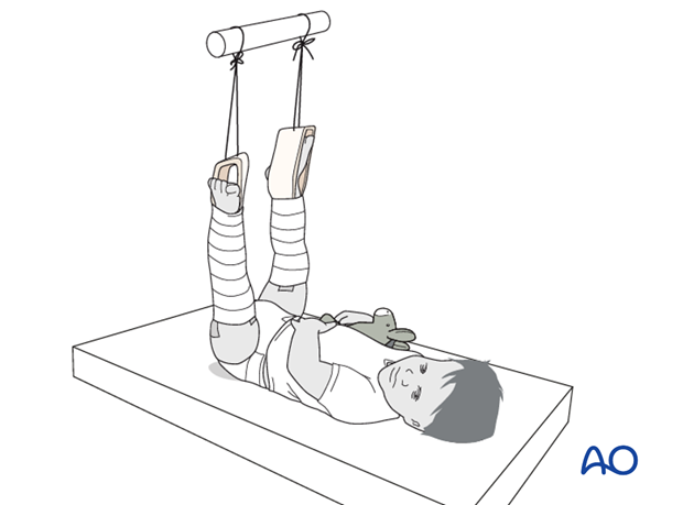
In children up to 2 years of age with a bodyweight up to 12–15 kg, overhead skin traction is preferable.
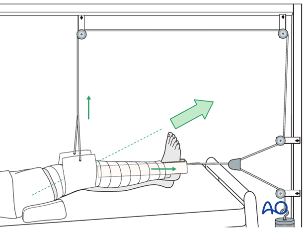
In children older than 2 years and heavier than 12–15 kg, longitudinal traction is an alternative. Traction can be applied with an adhesive bandage to the skin. Disadvantages include malunion and prolonged hospital stay.
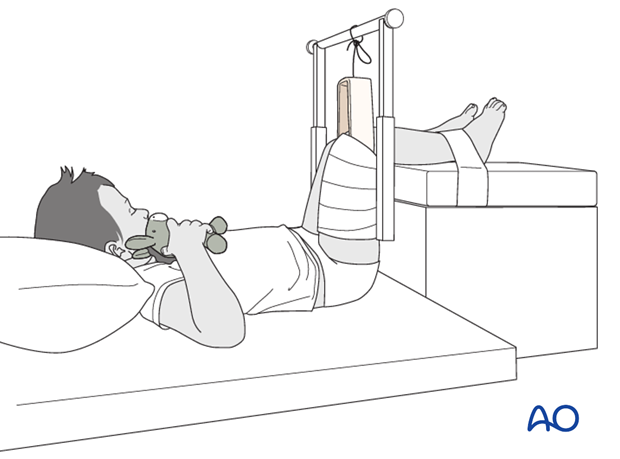
An alternative in children older than 2 years is 90/90 traction. Less weight is needed because of the support under the upper legs and fewer skin problems are therefore seen. A further advantage of this method is rotational control, which can be useful in unstable fracture patterns.
Preparation
Read the additional material on preoperative preparation.
2. Overhead skin traction
Position the patient supine on a bed.
Start with the uninjured side as this causes less pain.
Position the stirrup of the traction set with the strings for weight attachment at the foot leaving space for ankle movement.
Apply the long adhesive bandages from ankle to hip to distribute the skin tension forces over a large area to prevent blisters and skin necrosis.
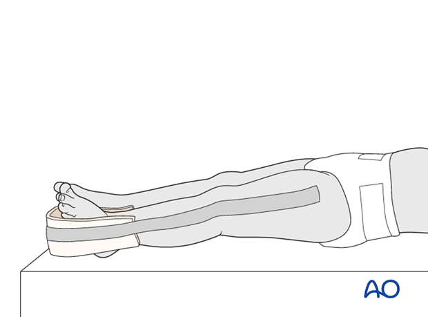
Wrap a bandage over the adhesive bandage to further reduce shear forces.
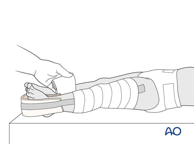
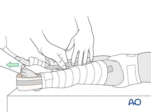
Use an assistant to apply gentle traction and provide stability when applying the bandage on the injured side.
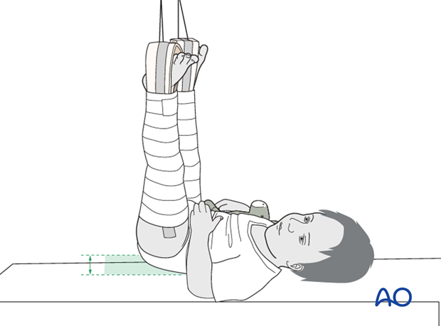
Position the hip in 90° flexion and use sufficient weight to raise the buttocks just above the sheet, producing the correct amount of traction on the leg.
Perform regular neurovascular observations and check for loosening of the bandage.
3. Longitudinal skin traction
Place the patient supine on the bed.
Position the stirrup of the traction set with the strings for weight attachment at the foot leaving space for ankle movement. Use an assistant to apply slight traction and provide stability when applying the bandage.
Apply the long adhesive bandages from ankle to hip to distribute the skin tension forces over a large area to prevent blisters and skin necrosis. Wrap a bandage over the adhesive bandage to further reduce shear forces.
Apply the sling just above the knee and place the leg on a support, eg a pile of towels, to produce 30° knee flexion.
This neutralizes the muscle forces, controls rotational deformities, and maintains the femoral bow, which may require a small pillow or towel under the thigh.
The resulting traction direction should be in line with the femoral axis.
The traction weight is determined by the age and weight of the child and should be sufficient to maintain length and alignment. 500 g per year of age is a good initial weight to use. The foot of the bed should be raised, tilting the bed, to stop the child being pulled down the bed.
Perform regular neurovascular observations and check for loosening of the bandage.

4. 90/90 traction
Place the patient supine with both lower legs on a support and the hip and knee flexed to 90°.
Apply an adhesive bandage to the entire upper leg on the injured side.
Adjust the lower leg support so that the buttocks are just lifted from the bed and stabilize the lower leg with a second adhesive bandage.

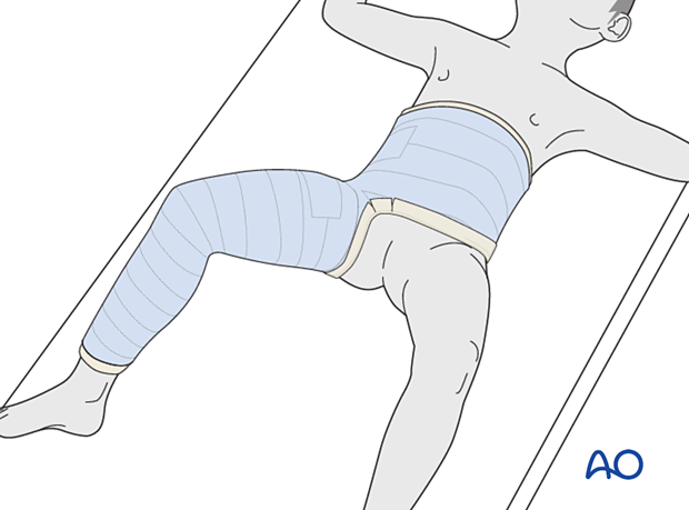
5. Delayed spica casting
Once the child is comfortable and moving or callus formation is visible on x-ray remove the traction and convert into a spica cast.
6. Removal of spica cast
The spica cast can be removed once healing is demonstrated on x-ray. This is age dependent and should generally be possible within 4–6 weeks.
7. Aftercare
Mobilization
Mobilization with crutches or a walker commences after completion of nonoperative treatment of the fracture.
Physical therapy may be required for gait training, hip and knee range-of-motion exercises and muscle strengthening.
Activities involving running, and jumping are not recommended for three months.

Follow-up
Clinical and radiological follow-up is usually undertaken after 2–4 weeks.
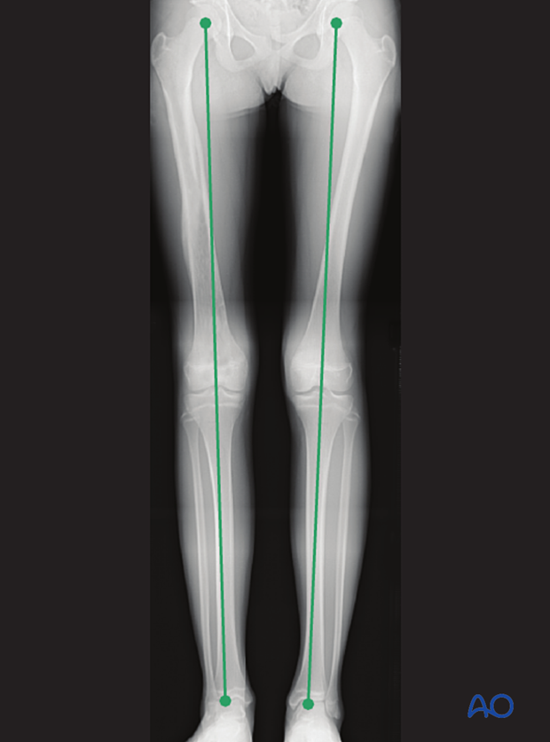
Clinical assessment of leg length and alignment is recommended at one-year. It uses a tape measure from the ASIS to the medial malleolus.
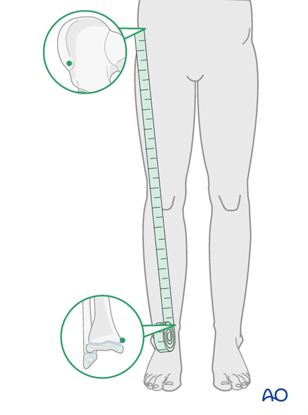
If there is any concern about leg length discrepancy or malalignment, long-leg x-rays are recommended.
Leg length is measured from the femoral head to the ankle joint.
