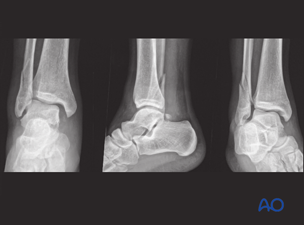
Radiological evaluation
1. Imaging of physeal injuries
Physeal injuries require radiological assessment to evaluate location, extent, displacement, and articular involvement. This will inform decision making, with a primary goal of anatomical reduction of the articular surface and physis, to minimize the risk of growth disturbance and joint degeneration.
Recommended reading:
- Nguyen JC, Markhardt BK, Merrow AC, et al. Imaging of Pediatric Growth Plate Disturbances. Radiographics. 2017 Oct;37(6):1791–1812.
2. Plain x-rays
Most fractures can be identified on plain x-rays.
AP and lateral views, centered at the ankle, are usually sufficient for diagnosis, but an x-ray that includes the proximal tibia and fibula may be necessary to exclude an additional proximal injury.
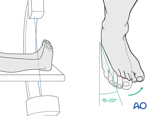
A mortise view may be necessary to evaluate articular and epiphyseal injuries.
The mortise view is an AP view of the ankle with 15°–20° internal rotation of the foot and ankle.
This provides a view of the lateral aspect of the distal tibial physis and is useful in identifying Tillaux fractures.
3. CT imaging
CT is indicated when further information about the fracture morphology is necessary for diagnosis and preoperative planning. This is often required for triplane and Tillaux fractures.
In children, fine cuts (2 mm) should be used for accurate definition of the fracture pattern.
CT is also used to assess the position and extent of bony bridge formation following physeal injuries.
4. MRI imaging
MRI is useful for identifying ligament and other soft-tissue injuries, chondral and physeal injuries.
5. Injury specific considerations
Salter-Harris I fractures
The undisplaced Salter-Harris I fracture is difficult to identify radiologically, and the presence of local tenderness is an important sign. An MRI may be necessary to confirm the presence of this injury.
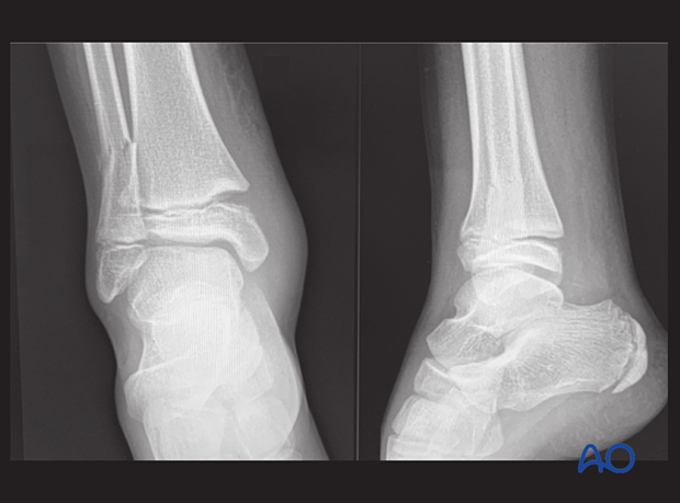
Tillaux fracture
A Tillaux fracture is a Salter-Harris III injury. It is characterized by a physeal fracture on the anterolateral corner of the distal tibia extending to the joint surface on the AP view.
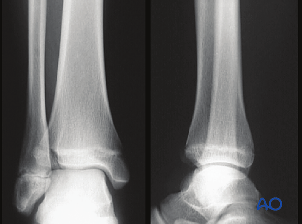
Triplane fractures
Triplane fractures will often consist of a Salter-Harris II injury on the lateral view and a Salter-Harris III injury on the AP or mortise view.
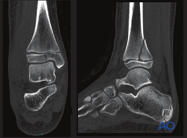
A CT scan will often provide information about the fracture configuration and displacement, including involvement of the articular surface.
The axial scans are also important in determining the trajectory of epiphyseal and metaphyseal screws.
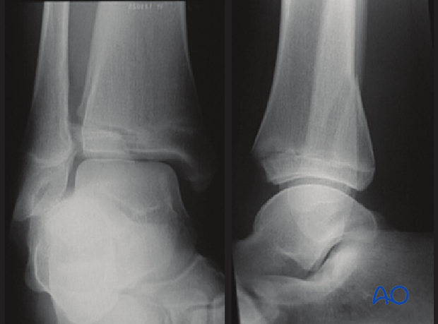
CT images are also useful to discriminate between two-, three-, and four-part triplane fractures.
Syndesmotic injuries
Divergence of the tibia and fibula, with disruption of the mortise on an AP x-ray indicates a syndesmotic injury.
