Safe zones for pin placement in the pediatric leg
1. Introduction
An ankle spanning external fixator requires insertion of pins into the tibial shaft and calcaneus.
These must avoid the physis and follow safe zones to reduce the risk of damage to the neurovascular structures.
2. Lower leg
Neurovascular structures
Common peroneal nerveThe common peroneal nerve runs laterally from the center of the popliteal fossa and curves distally around the fibular head. It divides into a superficial and deep branch. Injury to the deep branch will result in loss of ankle and toe extension.
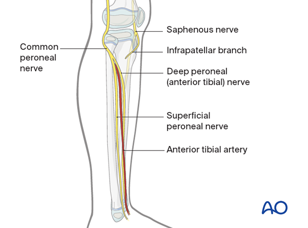
The saphenous nerve runs distally along the anteromedial aspect of the thigh. The infrapatellar nerve branches as it passes the knee joint.
There is no motor component, but an injury can cause cutaneous sensory loss in the infrapatellar region and medial aspect of the calf.
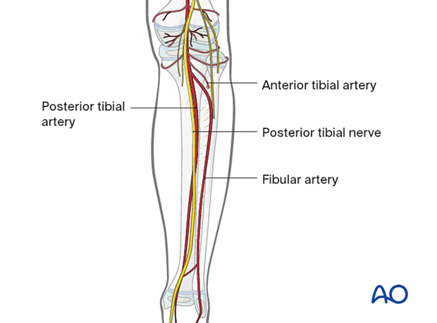
The popliteal artery runs through the center of the popliteal fossa. It separates into the anterior tibial artery, the fibular artery, and the posterior tibial artery at the level of the proximal tibial shaft.
Safe zone in the proximal third of the tibia
Pins should be inserted at least 2 cm distal to the physis to avoid growth disturbance.
Insert the pins on the subcutaneous surface or anterior crest as there is minimal muscle coverage and the risk of pin-track infection is, therefore, lower.
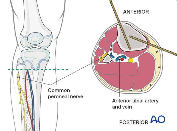
Safe zone in the midshaft of the tibia
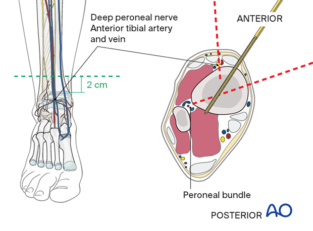
The anterior tibial artery and vein, together with the deep peroneal nerve, run close to the posterolateral border of the tibia.
These structures are at risk if a pin is inserted in the direction indicated by the red dotted line.
The pins should be inserted medial to the tibial crest, on the anteromedial aspect of the tibia, and are angled approximately 20° to the sagittal plane.
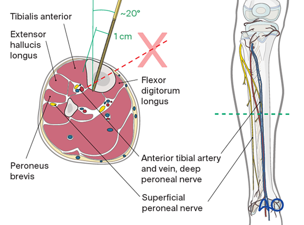
Safe zone in the distal third of the tibia
Pins should be inserted at least 2 cm proximal to the physis to avoid growth disturbance.
When inserting pins in the distal tibia, take into account the position of the anterior tibial artery and vein. Percutaneous insertion of pins in this area is dangerous. A 1 cm incision with dissection down to the bone will allow soft-tissue protection and safe insertion.
The peroneal bundle is located close to the posterolateral border of the tibia and is therefore at risk if pins are inserted in this direction.
A pin at this level should be inserted from anteromedial to posterolateral, as shown in the illustration. A second pin may be inserted from medial to anterolateral.

3. Foot
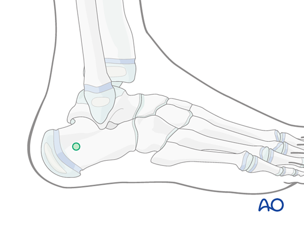
Pin placement in the calcaneus
The posterior tibial artery, medial and lateral plantar nerves, and the medial calcaneal nerve are at risk of penetration with a lateral calcaneal pin.
Locate the posterior tibial pulse as a guide to the path of the neurovascular bundle. If this is not palpable due to swelling, locate the pulse on the uninjured leg and use this to estimate the position on the injured side.
Use an image intensifier to avoid the calcaneal apophysis.
In larger children, the base of the fifth metatarsal may be used to place an additional pin.
