Splint immobilization without reduction
1. Introduction
Undisplaced fractures are immobilized for protection and comfort.
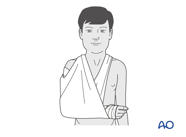
The recommended technique is to use a plaster or fiberglass backslab with 90° of elbow flexion.
A sling may be used for comfort according to the child's, or parental, preference.
2. Splint application
Application of cast padding
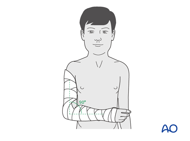
Cast padding is wrapped around the upper arm, elbow, forearm and hand, as far as the transverse flexor crease of the palm (the MP joints are left free). According to surgeon's preference a tubular bandage may be applied to the arm beneath the padding.
The elbow is held in 90° flexion and the forearm in neutral rotation.
It is important to make sure that the epicondyles of the humerus and the antecubital area are padded well.
Application of splint
A splint of fiberglass, or plaster, is applied on the posterior aspect of the arm and forearm. It should be wide enough to cover more than half the circumference of the arm and forearm.
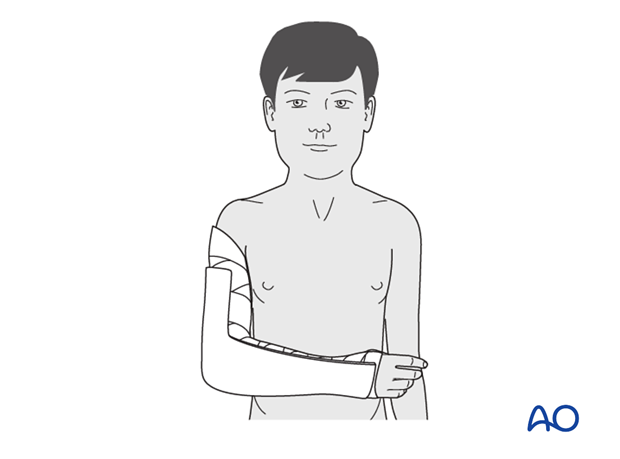
The splint is secured with a noncompressive bandage.
It is very important to ensure that this is not tight, so as to accommodate subsequent swelling.

Sling
The injured arm and cast are supported with a sling.

Analgesia
Nonnarcotic analgesia may be required.
Caregivers should be taught to monitor for excessive pain or other signs of potentially dangerous swelling.
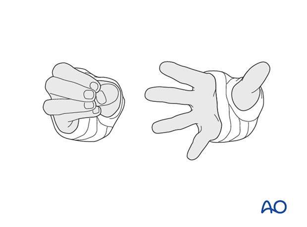
The ability to passively, or actively, fully extend the fingers without discomfort indicates normal perfusion and absence of neurological compromise, or muscle compartment compression.
3. Aftercare
Supracondylar humeral fractures heal rapidly and often within 3–5 weeks.
Compartment syndrome
Compartment syndrome is a possible early postoperative complication that may be difficult to diagnose in younger children.
The child should be examined frequently, to ensure finger range of motion is comfortable and adequate.
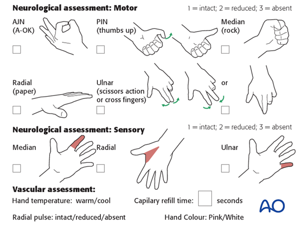
Neurological and vascular examination should also be performed.
Increasing pain, decreasing range of finger motion, or deteriorating neurovascular signs should prompt consideration of compartment syndrome.
Discharge care
When the child is discharged from the hospital, the parent/caregiver should be taught how to assess the limb.
They should also be advised to return if there is increased pain or decreased range of finger motion.
It is important to provide parents with the following additional information:
- The warning signs of compartment syndrome, circulatory problems and neurological deterioration
- Hospital telephone number
- Information brochure
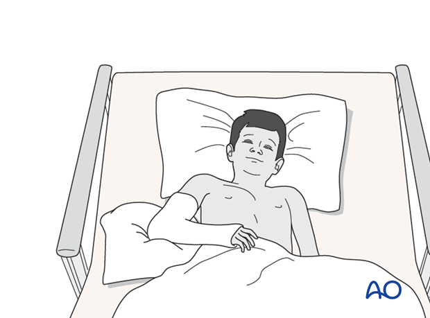
For the first few days, the elbow and forearm can be elevated on a pillow, until swelling decreases and comfort returns.
When the limb is comfortable, the child may optionally use a sling to support any splint if desired. Many children are more comfortable without a sling.
Follow-up x-rays
Control x-rays may be taken at one week following injury to assess fracture position. Further x-rays may be necessary at three weeks to assess fracture healing. This should be performed after the splint has been removed.
Removal of cast or splint
Splints or casts are usually removed within 3 weeks of the injury.
Recovery of motion
As symptoms recover, the child should be encouraged to remove the sling and begin active movements of the elbow.
The majority of elbow motion is recovered rapidly, usually within two months of splint removal. The older child may take a little longer.
Once the child is comfortable, with a nearly complete range of motion, he/she may resume noncontact sports incrementally. Resumption of unrestricted physical activity is a matter of judgment for the treating surgeon.


