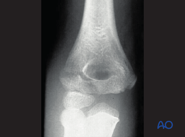
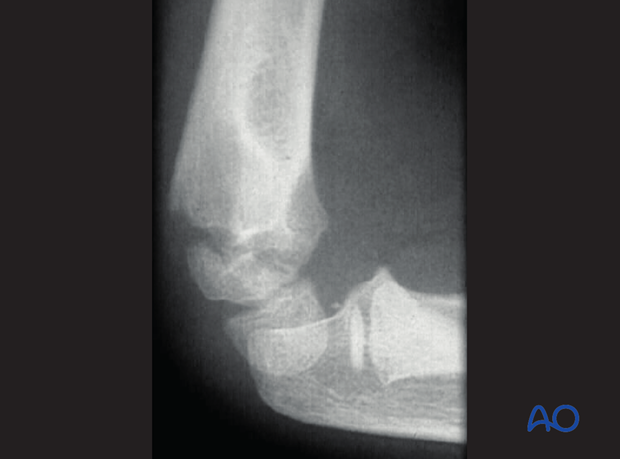
13-E/4.1L Simple epi-/metaphyseal, lateral condyle, SH IV
Salter-Harris IV fractures of the lateral condyle are the most frequent intraarticular fractures of the distal humerus in children.
The pattern of this fracture is characterized by a single epiphyseal fracture with a posterolateral metaphyseal wedge (Salter-Harris IV). There is a variation in the size of the metaphyseal fragment, depending on the anatomical alignment of the growth plate posteriorly. The classification is independent of fragment size and displacement.
The epiphyseal fracture may pass medial to the capitellar ossific center, or occasionally through it.
Another variation is in the initial stability of the fracture. Because of the thick epiphyseal cartilage around the capitellar ossification center, especially in younger children, the fracture at the level of the joint surface can be incomplete ("hanging fracture"). Hanging fractures can best be demonstrated by ultrasonography, or arthrography.
In these fractures there can be an associated injury of the medial collateral ligament. This can be evidenced by clinical tenderness over the damaged ligament. A medial injury is a strong indicator of elbow instability.
Other associated injuries include elbow dislocation, fractures of the radial head, or the medial epicondyle.
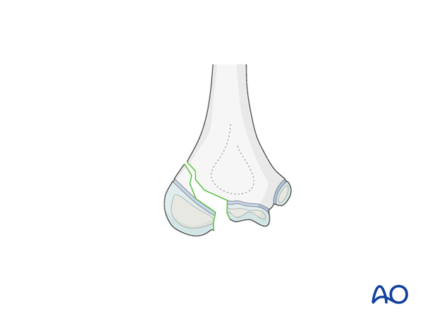
Radiologically this fracture can be difficult to diagnose if the child is very young or the fracture is minimally displaced. The fat pad sign is an important indirect indicator of an intraarticular lesion.
Internal oblique views are helpful for demonstrating the metaphyseal fragment.
Ultrasonography can also help to verify the diagnosis.
Arthrograms may be used when closed reduction is indicated (only for minimally displaced fractures).
MRI under sedation can sometimes be indicated in very young children.
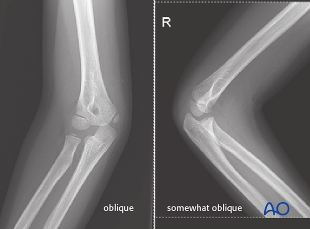
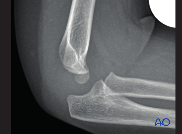
Careful examination of the lateral x-ray will often reveal a metaphyseal fragment not evident on the AP view.
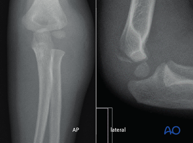
Very young child (under 3 years):
- In a very young child the diagnosis of such a fracture can be very difficult, especially in the AP view. Therefore, a true lateral view, or sometimes an oblique view, will show the posterior metaphyseal fragment
- This fracture is extremely rare in children younger than 2 years of age. It is important to be aware of the possibility of nonaccidental injury in this age group
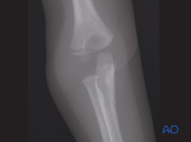
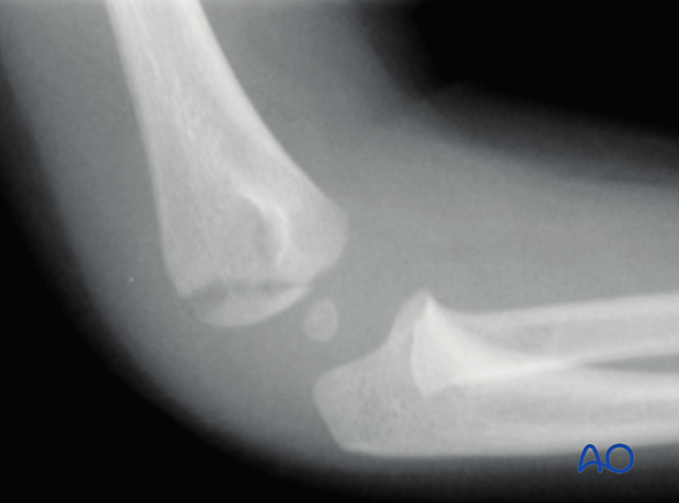
Very young child (under 3 years):
- In this patient, of similar age to the previous example, the fracture can be seen in the AP view, but not in the lateral view. These two cases demonstrate the importance of always obtaining both AP and lateral views for diagnosis
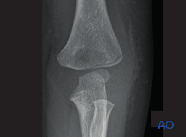
Older child (older than 6 years):
- In the older child, diagnosis of such a fracture is easier, as the metaphyseal fracture is usually visible in both AP and lateral views. If there is doubt, an oblique view should be taken
