Anatomy of the distal femur
1. Developmental anatomy
The epiphysis of the distal femur is present at birth. The physis at this location is responsible for 70% of the growth of the femur with almost 1 cm longitudinal growth per year.
It closes between 14 and 17 in females and between 15 and 19 in males with a wide variability in age.
2. Physeal anatomy
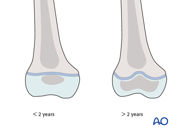
The physis is smooth and flat until the age of 2.
After this age, a central ridge and four quadrants of angulation develop and are responsible for the characteristic shape.
This shape is intrinsically stable and physeal displacement requires a larger transfer of energy than other anatomical sites.
The germinal zone is frequently damaged in displaced physeal fractures, causing growth disturbance.

3. Cartilage
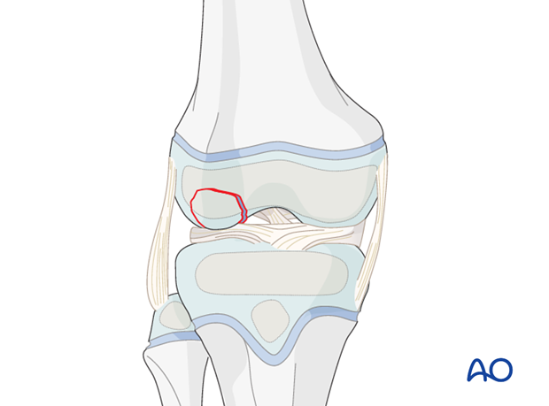
In skeletally immature patients, a large proportion of the distal femoral condyles are formed from epiphyseal or articular cartilage.
In contrast to predominately ligamentous injuries in adults, the distal femoral growth plate, epiphyseal cartilage or bone is more commonly involved in children but a combined injury may also occur.

4. Popliteal fossa
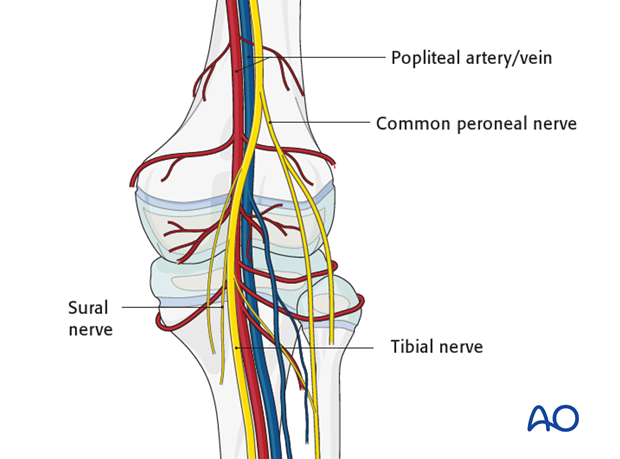
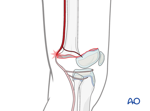
High energy distal femoral trauma is associated with damage to the neurovascular structures in the popliteal fossa.
Careful application of plaster casts is also important to avoid compromising these structures.

This is particularly relevant for displaced epiphyseal fractures of the distal femur.


