Medial approach to the pediatric distal femur
1. General considerations
The medial approach to the distal femur is most commonly used for open reduction of Salter-Harris II fractures. It can also be used to expose intraarticular fractures, a Hoffa-type fracture, osteochondroma, or a neoplastic lesion of bone.
It also provides access to the neurovascular bundle when a distal femoral fracture is complicated by an arterial injury.
The approach can be extended to expose the posterior cruciate ligament.
This approach also allows limited access to the posterior aspect of the distal femur.
2. Open reduction of Salter-Harris II fractures
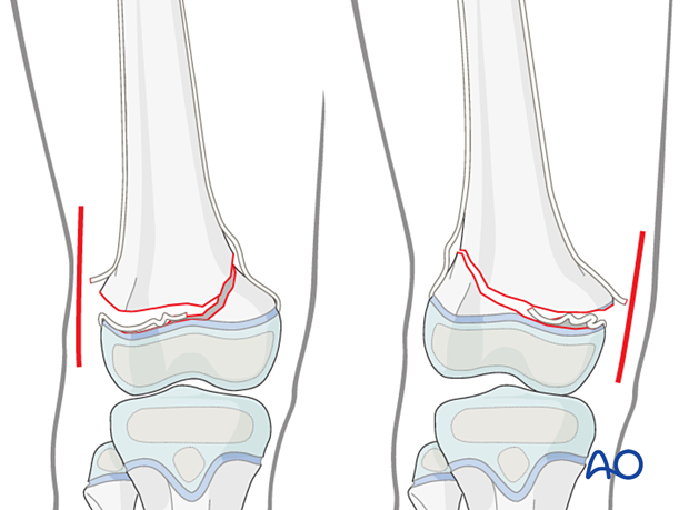
This approach is commonly used if closed reduction of a Salter-Harris II fracture has failed.
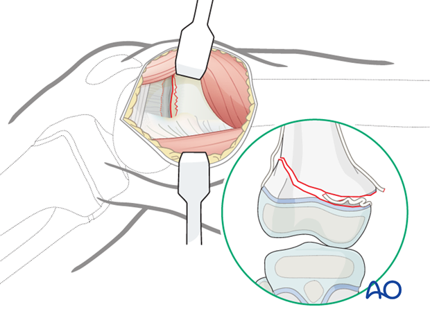
In this case the block to reduction is almost always periosteum entrapped in the fracture on the side that has failed in tension. The initial approach should be made from this side.
This means making the initial incision on the side where the shaft fragment is prominent. This is opposite to the side with the metaphyseal (Thurstan Holland) fragment.
3. Skin incision
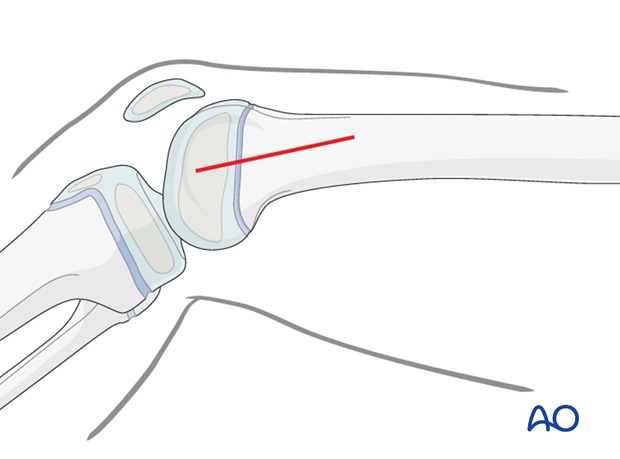
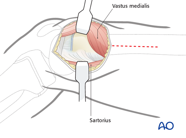
Perform a straight-line incision along the posterior border of the vastus medialis. The incision can be extended as far proximally as needed.
Exposure of the growth plate can be accomplished through a 4–5 cm incision beginning at the joint line and extending proximally to expose the physis and distal metaphysis.

4. Deep dissection for physeal fractures
Identify the distal and posterior edge of the vastus medialis, incise and mobilize the muscle.
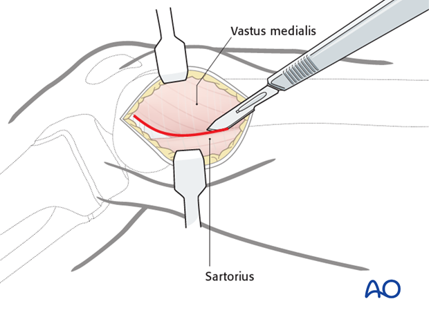
Retracting the vastus medialis anteriorly gives good visualization of the growth plate and distal metaphysis without further soft-tissue dissection.
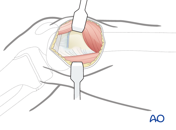
Look for interposed periosteum attached to the distal epiphyseal fragment.
5. Proximal extension
Identify the anterior edge of the sartorius.
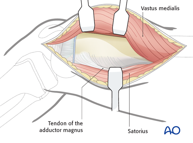
To aid the dissection, flex the knee, to allow the anterior border of the sartorius to be retracted posteriorly. This will allow exposure of the tendon of the adductor magnus. The adductor magnus tendon inserts into the adductor tubercle anteriorly.
6. Exposure
Retract the adductor magnus and its tendon posteriorly and retract the vastus medialis anteriorly to expose the femur.
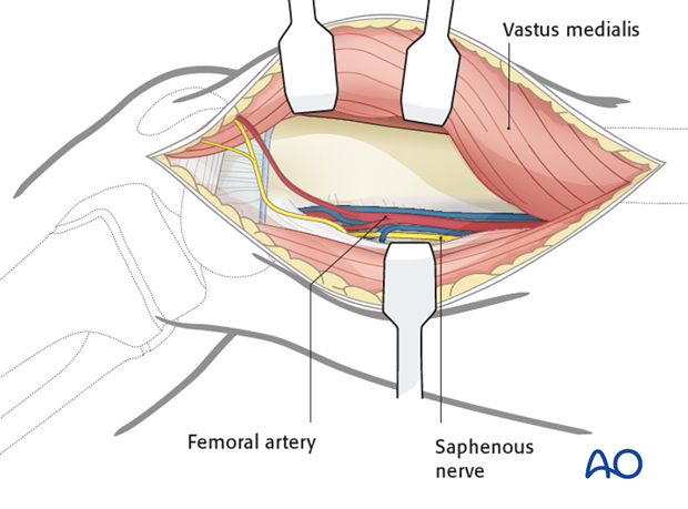
The popliteal neurovascular bundle lies in the popliteal space posterior to the femur and, if necessary, can be exposed through this approach. This is facilitated by blunt dissection deep to adductor magnus.
A capsulotomy can be performed if inspection of the joint surface is needed.
7. Wound closure
After careful hemostasis, close the skin and subcutaneous tissues in a routine manner.


