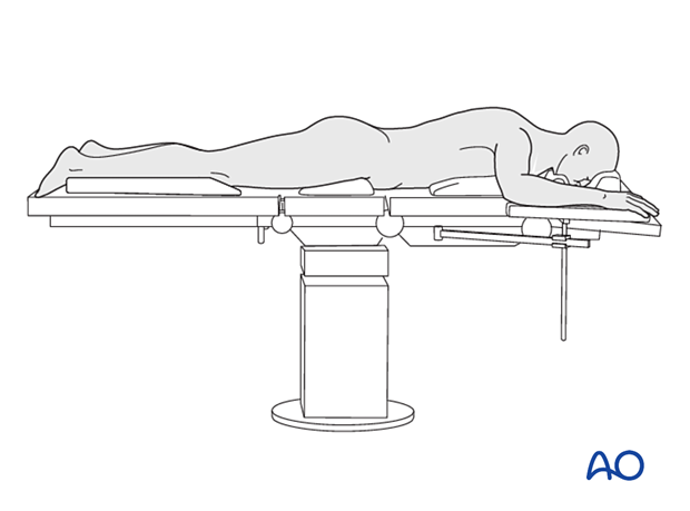
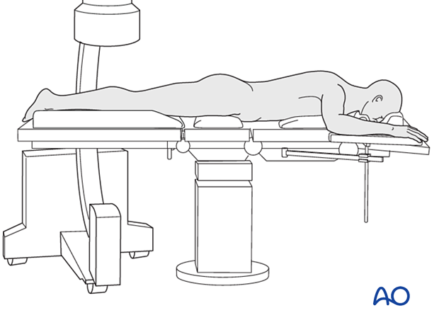
Prone position
1. Patient positioning
The patient is positioned prone on a radiolucent table or a standard table with a radiolucent extension. Rolls are placed underneath the thorax and pelvis to allow for optimal ventilation. All bony prominences are padded to prevent pressure sores.
The face is protected with a cushion to avoid pressure on the eyes.
The injured leg is elevated with a bump to facilitate lateral imaging with the C-arm.
If desired, apply a tourniquet around the upper thigh.
Prepare the entire leg circumferentially, from the toes to the upper thigh. Drape so the leg is completely mobile.
Note: Patients with unstable spine fractures, chest injuries, etc, have increased risks associated with prone positioning. Consider carefully whether these risks are worth taking.
Note: Improve the preoperative preparation with the WHO Surgical Safety Checklist.

2. C-arm placement
C-arm positioning must allow for perpendicular (AP and lateral) images of the knee, entire tibia, and ankle. The C-arm should be placed on the non-injured side of the patient.


