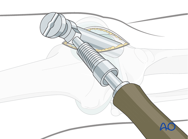
Semi-extended lateral parapatellar approach for intramedullary nailing of the tibial shaft
1. Determination of the entry point
In the frontal plane, the entry point is in the medial aspect of the lateral tibial spine. It is typically located in line with the medullary canal (3 mm medial to the tibial crest). In the sagittal plane, the entry point should be located just distal to the angle between the tibial plateau and the anterior tibial metaphysis, at the edge of the anterior joint line.
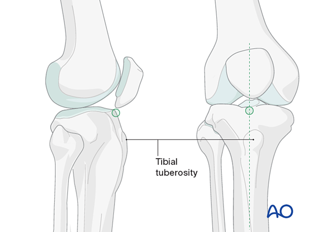
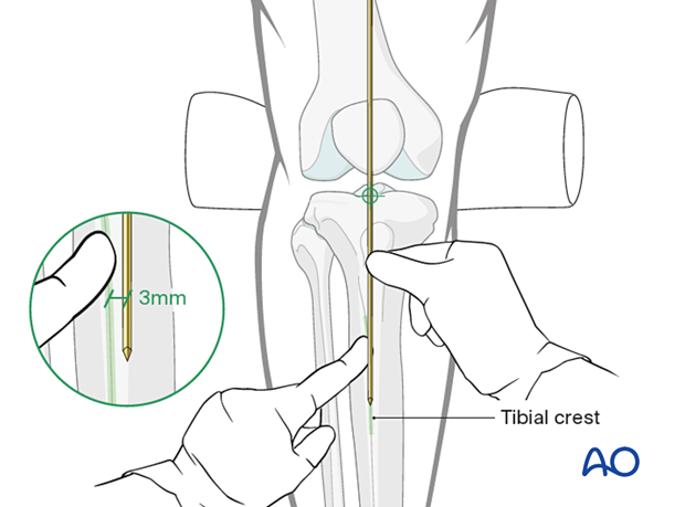
To find the correct entry point, identify the tibial crest and place a guide wire along it, extending proximally over the knee.
The correct insertion point will be at the intersection of the guide wire and the tibial plateau.
Use the image intensifier with proper AP and lateral images of the knee and proximal tibia to confirm proper guide wire placement.
2. Skin incision
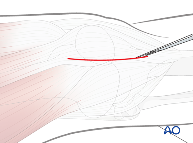
Make a longitudinal skin incision over the patella, or a longitudinal incision over the lateral border of the patella. This incision should allow access for performing a lateral parapatellar arthrotomy.
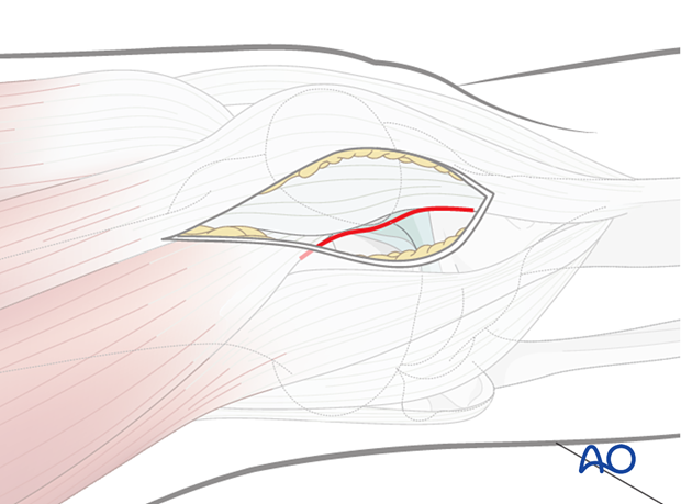
The image shows a laterally-based incision.
3. Parapatellar incision
The incision must go through the lateral parapatellar area so as to allow the guide sleeves for the tibial nail to be inserted as atraumatically as possible onto the anterior tibial crest. Care must be taken not to damage the articular surfaces of the patella and distal femur. In some instances, depending on the mobility of the patella, the incision of the parapatellar retinaculum must be increased to protect the articular cartilage. The joint capsule is usually not opened in this approach.
Divide the retinaculum leaving a cuff of tissue of a few millimeters on the patella for later repair.
Extreme care must be taken with insertion of the sleeve, and an attempt should be made to keep it inserted throughout the entire case to protect the cartilage.
