Infrapatellar approach for intramedullary nailing of the tibial shaft
1. Determination of the entry point
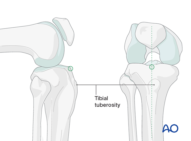
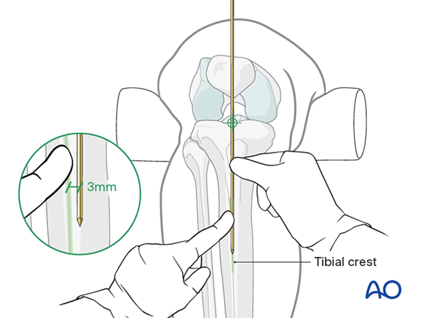
In the frontal plane, the entry point is in the medial aspect of the lateral tibial spine. It is typically located in line with the medullary canal (3 mm medial to the tibial crest). In the sagittal plane, the entry point should be located just distal to the angle between the tibial plateau and the anterior tibial metaphysis, at the edge of the anterior joint line.

To find the correct entry point, identify the tibial crest and place a guide wire along it, extending proximally over the knee.
The correct insertion point will be at the intersection of the guide wire and the tibial plateau.
Use the image intensifier with proper AP and lateral images of the knee and proximal tibia to confirm proper guide wire placement.

2. Skin incision
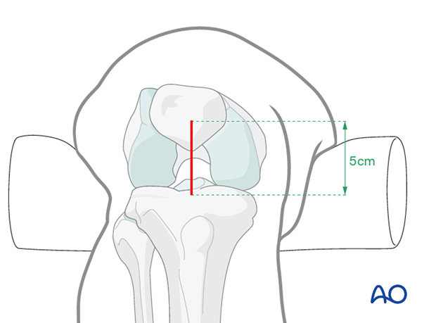
Make a longitudinal skin incision over the planned entry point. Extend it 3–5 cm proximally from the level of the tibial plateau.
An incision that is too medial interferes with proper entry into the medullary canal.

3. Tendon incision
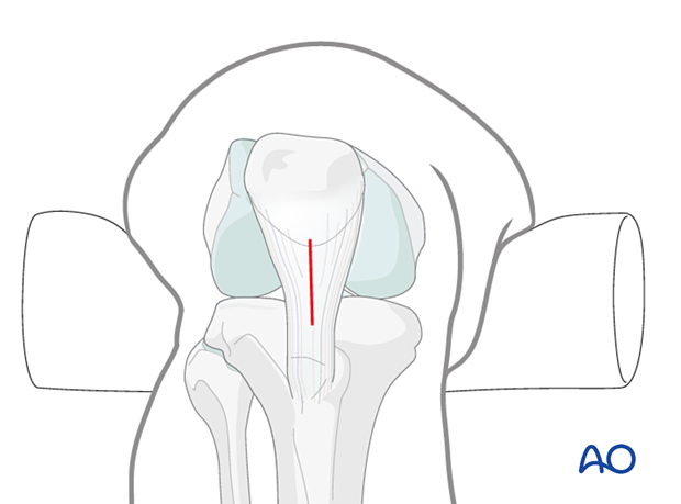
The incision goes through the patellar tendon. It is important that it is made directly in line with the medullary canal.
The patellar tendon is incised proximally from the patella bone and distally until the proximal tibia is appropriately exposed.
Proximal tibial shaft fractures tend to develop a valgus deformity if the nail entry site is placed too medially. A carefully placed skin incision aids in proper guide wire positioning, and therefore proper nail positioning.
Studies have found no difference in knee pain between an incision through or beside the patellar tendon.


