Approach to the posteromedial surface of the tibia
1. Indications
The posteromedial approach can be used for open plate fixation of the tibia on its posterior surface. Typically, this approach would be chosen, when direct exposure for ORIF is desired, but only the posteromedial soft tissues are safe to incise.
Note that this incision is also the one that might be used for a medial fasciotomy for compartment decompression.
The posterior surface of the tibia is relatively flat and therefore little contouring of the plate is necessary.
This approach allows access only to the tibia, not the fibula.
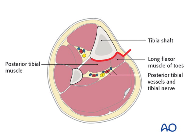
2. Positioning
The patient may be positioned either supine or prone. When positioned supine, the patient’s leg must be flexed at the knee and externally rotated at the hip to provide good exposure. In the prone position, the leg requires less manipulation.
3. Anatomy
Triangular shape of the tibia
The lateral and posterior surfaces of the tibia are covered by muscle. The anteromedial surface has only a thin layer of subcutaneous tissue and skin. This surface provides less blood supply to the underlying bone.
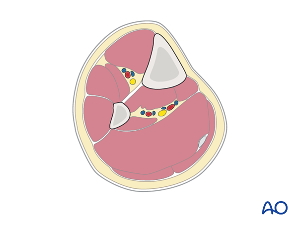
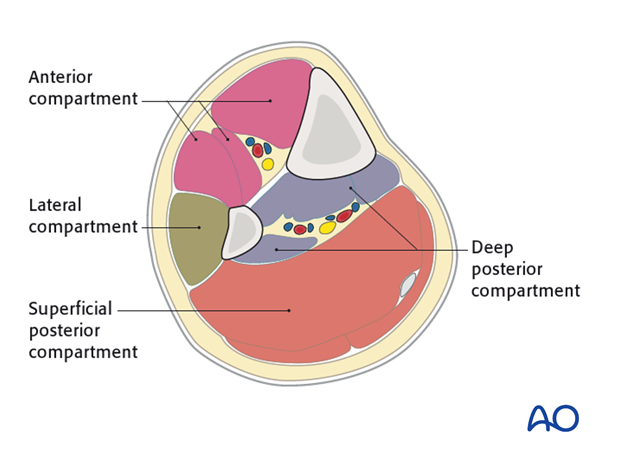
The lower leg has four compartments:
- Anterior
- Lateral
- Deep posterior
- Superficial posterior
The anterior compartment has three muscles and one main artery and nerve: Tibialis anterior, extensor hallucis longus, extensor digitorum longus; the anterior tibial artery and deep peroneal nerve.
The lateral compartment has two muscles and one nerve. The muscles are the peroneus longus and brevis and the superficial peroneal nerve.
The deep posterior compartment has three muscles and two arteries and one nerve: The muscles are the tibialis posterior, the flexor hallucis longus and the flexor digitorum longus. It also has the peroneal artery and the posterior tibial artery as well as the tibial nerve.
The superficial posterior compartment has just two muscles in it: The gastrocnemis and soleus muscles and the sural nerve.
4. Skin incision
The length of the incision varies according to fracture location and selected plate. The posteromedial boarder of the tibia is first palpated throughout its length. The incision is then made in parallel 1-2 cm posterior to the posterior tibial boarder.
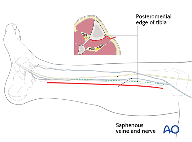
5. Dissection
Subcutaneous dissection follows carefully so as to identify and/or protect the saphenous vein and nerve. These are typically mobilized anteriorly.
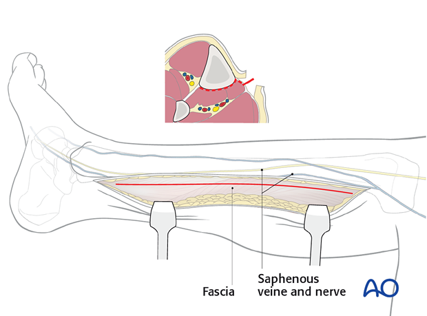
The fascia is then incised in line with the skin incision and the superficial and deep posterior compartments are mobilized. Gastrocnemius, soleus and flexor digitorum will be identified and mobilized with a soft-tissue elevator, depending on the level of the fracture.
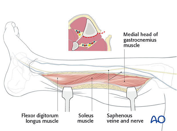
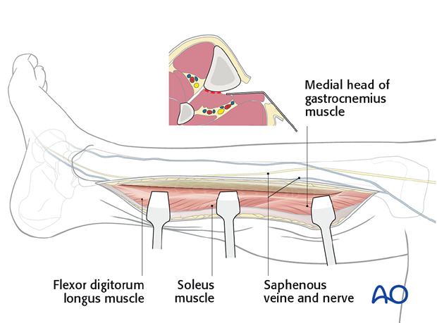
The dissection should be done in an extra-periosteal plane.
Dissection is not necessary beyond the posterolateral aspect of the tibia.
In this way, the middle 3/5 of the posterior tibia can be effectively exposed.
6. Wound closure
The deep fascia should almost always be left open to minimze the risk of a compartment syndrome affecting the deep posterior space. Only the skin and subcutaneous layers are closed.


