Approach to the anterolateral surface of the tibial shaft
1. Indications
The anterolateral approach, through an incision slightly lateral to the tibial crest, reflects the anterior compartment muscles from the lateral tibial surface.
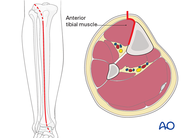
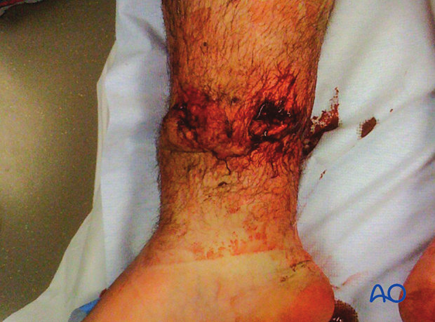
This approach is ideal for use when the medial soft tissues are compromised, such as with open fractures, as illustrated, where the wound overlies the site for a medial plate.
The disadvantage of this approach is that the exposure mobilizes the muscles of the anterior compartment, thus compromising tibial blood supply.
2. Anatomy
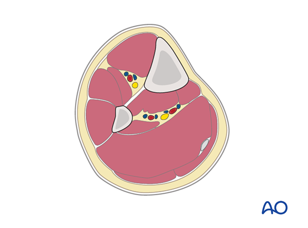
The lateral and posterior surfaces of the tibia are covered by muscle. The anteromedial surface has only a thin layer of subcutaneous tissue and skin. This surface provides less blood supply to the underlying bone.
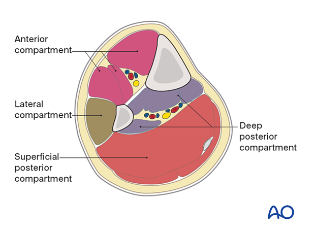
The lower leg has four compartments:
- Anterior
- Lateral
- Deep posterior
- Superficial posterior
The anterior compartment has three muscles, one main artery, and one nerve: the tibialis anterior, extensor hallucis longus, extensor digitorum longus, the anterior tibial artery, and the deep peroneal nerve.
The lateral compartment has two muscles and one nerve: the peroneus longus and brevis, and the superficial peroneal nerve.
The deep posterior compartment has three muscles, two arteries, and one nerve: the tibialis posterior, flexor hallucis longus and flexor digitorum longus, and the peroneal and posterior tibial arteries, as well as the tibial nerve.
The superficial posterior compartment has two muscles and one nerve: the gastrocnemius, the soleus, and the sural nerve.
3. Skin incision
A longitudinal incision lies 1–2 cm lateral to the tibial crest and continues distally towards the ankle joint along the line of the anterior tibial tendon.
The length of the incision depends on the plate length.
The distal extension of the anterolateral approach is helpful for distal tibial fractures but is obstructed by the muscles and neurovascular structures of the anterior compartment.

4. Dissection
The fascia is incised just lateral to the tibial crest, and the dissection is carried down extraperiosteally along the lateral surface of the tibia.
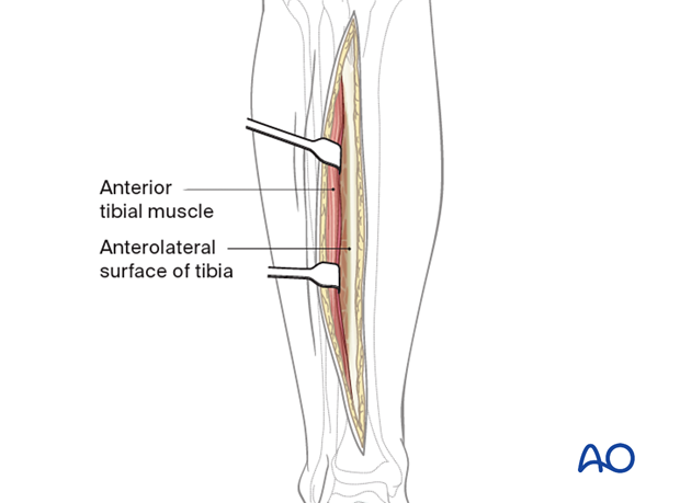
The periosteum is left intact, though it may require mobilization near the fracture site in order to expose fracture edges. This is commonly done in preparation for direct anatomical reduction.
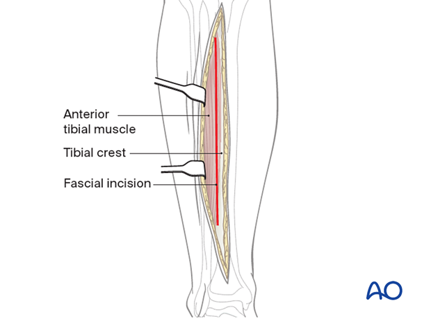
Retraction of the anterior tibial muscle should be limited, to show only the essential part of the anterolateral surface of the tibia.
Near the junction of the middle and lower thirds of the tibia, the anterior tibial artery and the deep peroneal nerve come together and approach the lateral tibial surface. They wrap obliquely anteriorly and distally around the tibia. In the distal metaphyseal area, they lie on the periosteum, under the myotendinous portion of the anterior tibial muscle, extensor hallucis longus, and extensor digitorum longus. If the exposure extends into the distal third of the tibia, the surgeon should identify and protect the neurovascular bundle. With care, it can be mobilized from the tibial surface, along with the anterior compartment muscles. This makes it possible to pass a plate more distally on the anterolateral surface, all the way to the distal end of the tibia, if necessary.
5. Wound closure
Only the skin and subcutaneous tissues should be closed. The fascia should be left open. This is important to minimize the risk of compartment syndrome.


