Cerclage compression wire
1. Principles
Anatomy
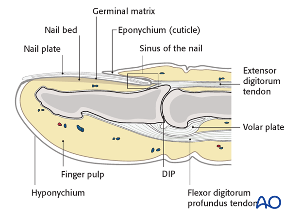
The distal phalanx is divided into three anatomical parts: most proximally, the metaphysis, followed by the diaphysis (“shaft”), and finally the ungual tuberosity (“tuft”).
The base of the distal phalanx has a prominent dorsal crest at the insertion of the extensor tendon. The tendon is also adherent to the distal interphalangeal (DIP) joint capsule.
On the palmar surface is the insertion of the flexor digitorum profundus tendon. This is also adherent to the volar plate.
The flexor tendon inserts into the whole width of the base of the distal phalanx.
The volar plate is very flexible, allowing hyperextension of the DIP joint and pulp-to-pulp pinch.
The vascularity of the extensor tendons is more precarious than that of the flexor tendons. This prolongs extensor tendon healing time.
Diagnosis
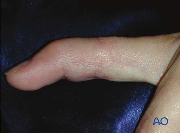
Discontinuities of the extensor insertion are often referred to as “mallet injury” or “baseball finger”. They can be purely tendinous, or bony avulsion fractures.
Diagnosis is based on
- the clinical history of the trauma,
- deformity, pain and swelling located in the dorsal aspect of the DIP joint,
- inability actively and fully to extend the DIP joint,
- x-rays.
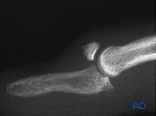
AP and true lateral x-rays of the DIP joint are necessary for the diagnosis of fracture avulsions.
Low-energy radiographs, as used to visualize soft tissues, can be useful in identifying small flakes of bone.
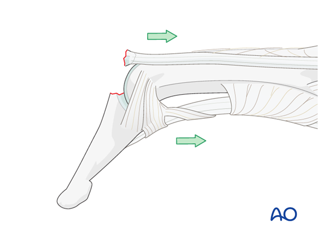
Flexion injury
The commonest cause of these injuries is forcible flexion of the actively extended DIP joint, as when stubbing a straight finger against resistance.
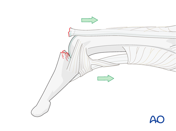
Axial compression injury
Occasionally, the injury is the result of an axial overload of the terminal segment of the finger, causing joint impaction and a dorsal marginal fracture, which is retracted by the pull of the extensor tendon.
Fracture subluxation of DIP joint
An obliquely orientated axial compression force sometimes results in a dorsal marginal fracture, involving approximately half the articular surface, and can disrupt the collateral ligaments.
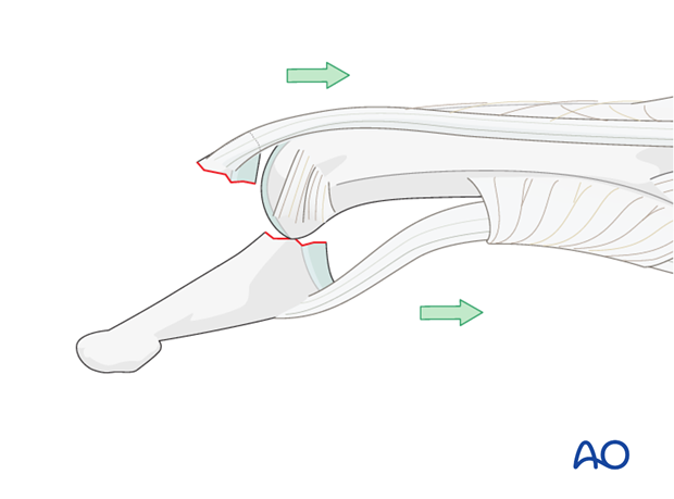
In such instances the pull of the flexor digitorum profundus results in palmar subluxation of the distal phalanx.
This injury represents a strong indication for ORIF.
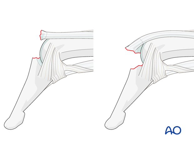
Spectrum of injury – partial disruption
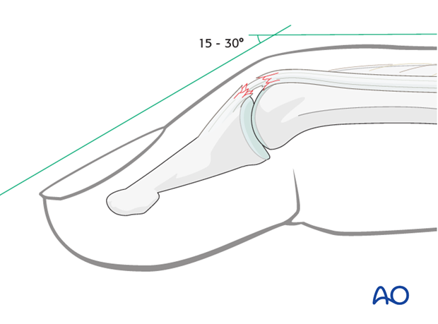
These injuries may be complete disruptions or partial disruptions.
The nature of the disruption may be either a tear of the tendon insertion without fracture, or a bony avulsion of the tendon from the dorsum of the base of the distal phalanx, the bony fragment being of variable size.
In incomplete tendon injuries the resulting extension lag is no greater than 30 degrees. The patient retains a partial ability actively to extend the DIP joint.
In complete disruption of extensor mechanism, the patient is unable actively to extend the DIP joint.
The flexor digitorum profundus exerts a flexion deforming force onto the distal phalanx, partly counterbalanced by the intact oblique retinacular ligaments and the collateral ligaments.
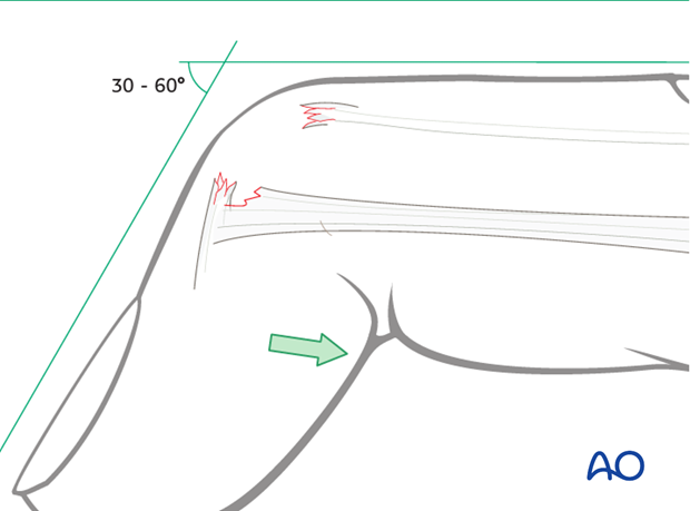
A similar clinical picture is presented by bony avulsion of the extensor mechanism at its insertion. The dorsal avulsion fracture is of variable size.
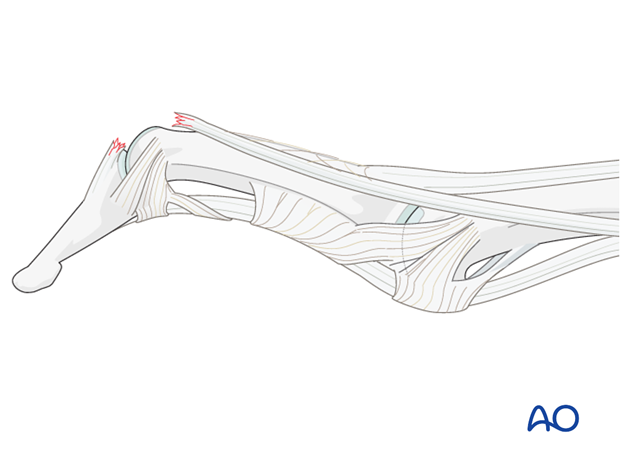
Swan-neck deformity
In some patients, elasticity of the ligaments and a lax PIP joint can result in swan-neck deformity, because after disruption of the extensor mechanism at the DIP joint, all extensor forces are concentrated on the PIP joint.
Nonoperative treatment (splinting)
Nonoperative treatment can produce excellent results, with near-normal range of motion, due to bone remodelling, even in the presence of persisting fragment displacement.
Nonoperative treatment is based on immobilization of the DIP joint in extension, leaving the PIP joint free.
Duration of immobilization
The DIP joint should be immobilized in extension for 8 weeks. It must be impressed on the patient that splintage should be uninterrupted during this period.
It must be kept in mind that the vascularity of this area is precarious, even in healthy patients, and healing is slow. Any shorter period of immobilization risks re-rupture. Joint stiffness is unusual after these injuries.
Result
Frequently, patients may complain of a dorsal prominence, and of loss of the last 10-20 degrees of extension of the DIP joint. Dorsal tenderness rarely persists for more than a month or so after removal of the splint.
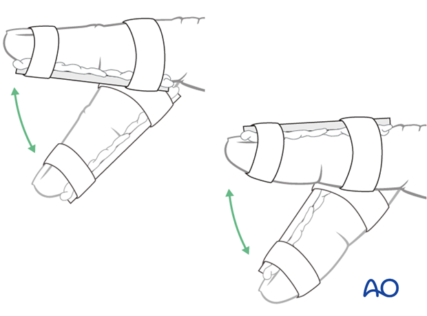
Nonoperative treatment (splinting)
Dorsal splint vs palmar splint
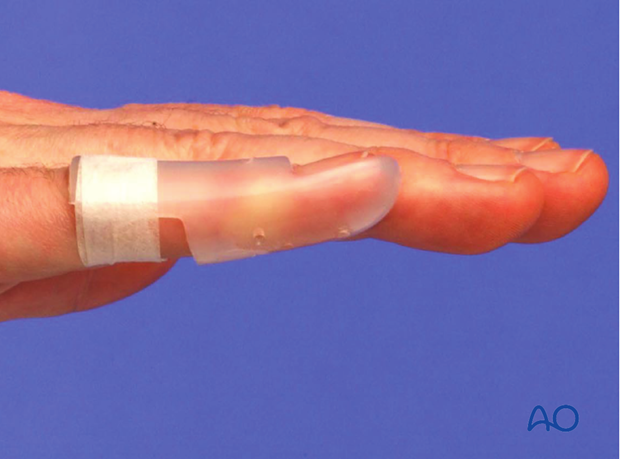
Using a dorsal splint has the advantage of leaving the patient with the ability to pinch while the digit is immobilized.
However, proponents of palmar splintage argue that the palmar aspect is better cushioned than the dorsal and thereby can tolerate the splint better.
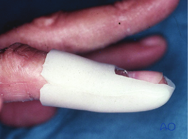
Contoured custom thermoplastic splint
The advantage of a custom thermoplastic splint is that it is better adapted to the shape of the finger, and easier to change.
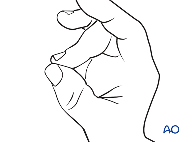
Cleaning
Instruct the patient to keep the finger in extension by pinching it with the thumb when the splint is taken off for cleaning.
Flexing the finger may delay the healing process.
Pitfalls of nonoperative treatment
Hyperextension of the DIP joint
Do not attempt to immobilize the joint in hyperextension. This can compromise the precarious vascularization of the skin of the dorsal aspect of the joint and might provoke ischaemia and possible necrosis.
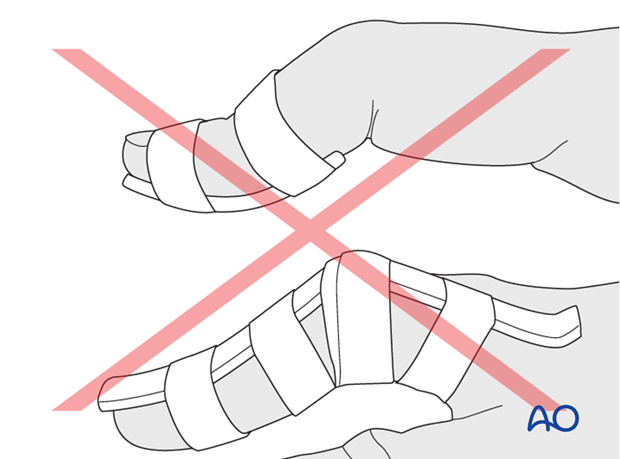
Immobilization of the PIP joint
Do not immobilize the PIP joint as this may result in flexion contraction of the joint.
2. Operative treatment: caveat
Inexperienced handling of this area may harm the germinative matrix of the nail and cause permanent deformity. Consider that nonoperative treatment is almost always a viable alternative in these fractures, often with comparable results. Operative treatment should only be attempted by experienced hand surgeons, in selected cases. Absolute indications for surgical intervention are:
- Open fractures
- Palmar subluxation of the DIP joint
It is wise always to use magnifying loupes in these surgical procedures.

3. Approach
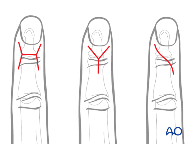
For this procedure a dorsal approach to the DIP joint is normally used.
4. Reduction
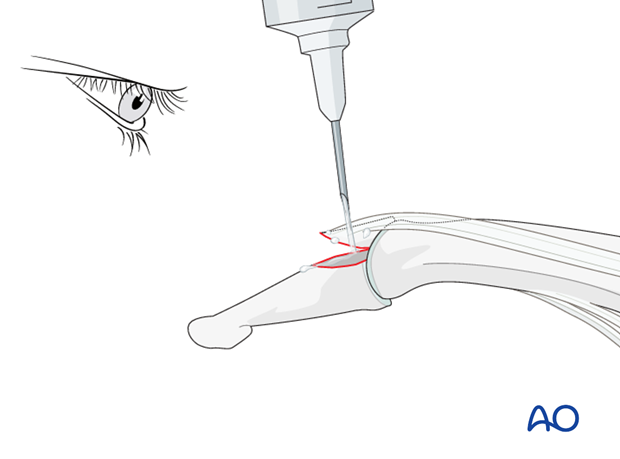
Clean the fracture site
Flex the DIP joint. In order to gain better visualization of the fracture, use a syringe to clean out blood clots with a jet of water.
Often the degree of comminution is not apparent from the x-rays, and can only be determined under direct vision.
Use a dental pick to carefully free interposed tissues, and to remove blood clots and other debris.
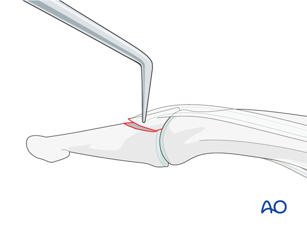
Reduce the fracture
Extend the DIP joint. Put manual pressure on the palmar side of the distal phalanx.
Now complete the reduction with help of a dental pick.
5. Wire fixation
Prepare transverse hole
Use a drill guide and a 1.5 mm drill bit to drill a transverse hole in the diaphysis of the distal phalanx.
The drill hole must be located dorsal to the mid-axis of the distal phalanx.
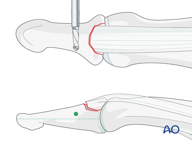
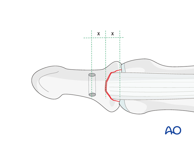
The location of the drill hole should be the same distance from the fracture line as the avulsed fragment’s length (usually approximately 5 mm). This will help the wires to cross above the fracture line, providing optimal force distribution.
Pass wire through the hole
Pass a 0.6 mm stainless steel wire through the hole. Alternatively, a 4.0 nonabsorbable multifilament suture can be used, double-mounted in a straight needle.
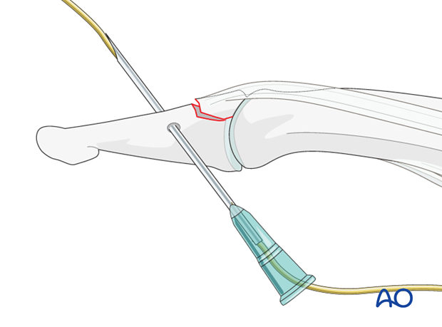
In order to preserve the delicate soft tissues, use a 16 hypodermic needle to thread the wire through the drill hole, and a hemostat to retrieve it.
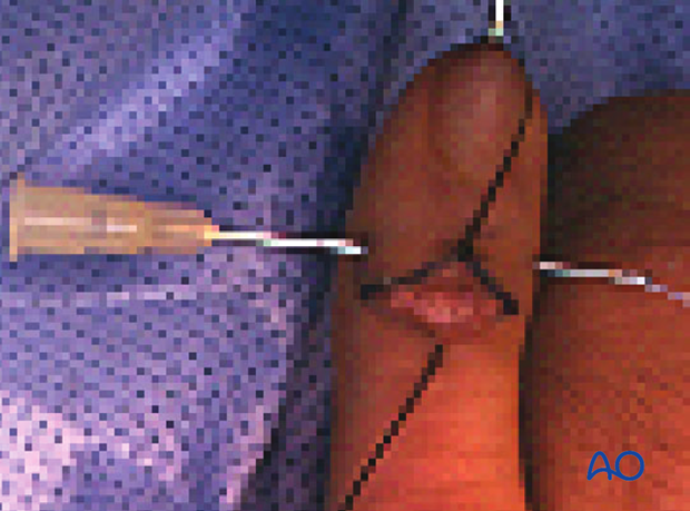
Pass wire through terminal extensor tendon
Pass the wire through the terminal extensor tendon, as close as possible to its insertion into the bone, using a 16 hypodermic needle.
Do not place the wire on top of the terminal extensor tendon, as this may cause necrosis by pressure.
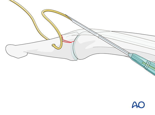
Tighten the wire
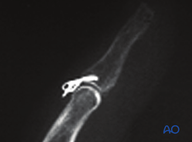
Now make a figure of eight and tie the wire.
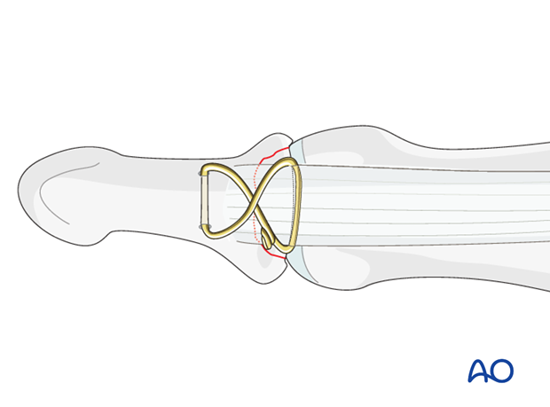
Check reduction using image intensification.
Tighten the wire.
Pitfall: overtightening
Do not overtighten the wire, as the terminal extensor tendon may be cut, or the bone penetrated, loosening the fixation.
Avoid soft-tissue irritation
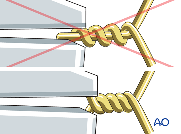
Cut the wire and bend it along the phalanx in order to avoid soft-tissue irritation.
When tightening the wire, ensure that both ends are twisted around each other rather than twisting one end around the other straight end.
Pitfall: tilting
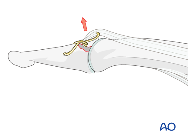
Depending on the fracture geometry, the avulsed fragment may tilt when the wire is tightened.
If this is the case, and the fracture size allows it, a 0.6 mm K-wire can be inserted to secure the reduction.
Alternative: use K-wire in avulsed fragment
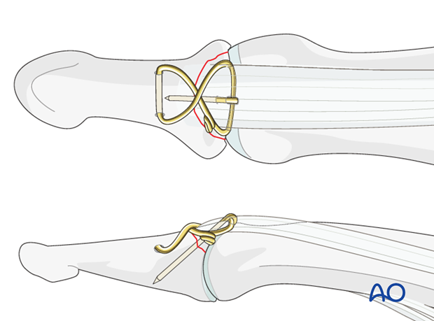
If the avulsed fragment is large enough, a 0.6 mm K-wire can be inserted to secure it to the main fragment of the middle phalanx. This K-wire can then be used to anchor the cerclage compression wire on the avulsed fragment.
Note: We now prefer the term “Cerclage compression wire”. This was previously referred to as a “Tension band wire”.
Check palmar protrusion of the K-wire using image intensification. Then retract the K-wire by about 2 mm, bend it through 180 degrees, and impact it into the bone. The K-wire must engage the far cortex, but should not protrude beyond.
The K-wire will prevent tilting of the fragment after tightening of the wire.
6. Alternative fixation: suture anchors
Indication
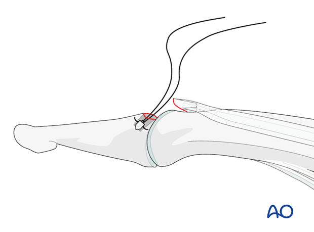
In very small fragments, suture anchors represent a good alternative.
Depending on the size of the fragment, two anchors can be used.
Fixation
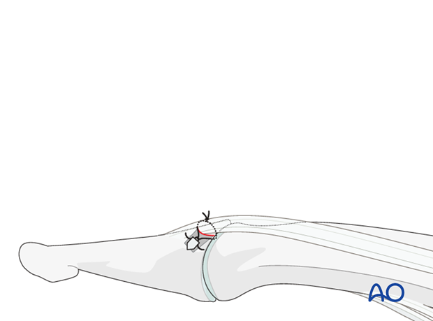
Use an anchor that has approximately the same breadth as the avulsed fragment.
Use 4.0 multifilament nonabsorbable sutures. Tie the sutures around the avulsed fragment.
7. Aftertreatment
Aftertreatment depends on the size of the fragment, the quality of the bone, and the stability gained by the fixation.
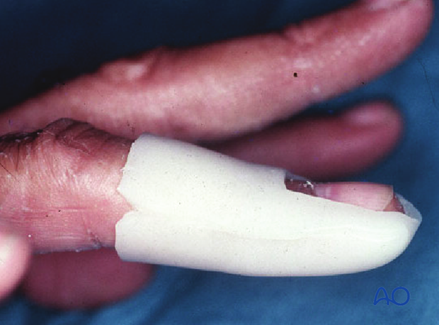
The DIP joint is immobilized in extension in a palmar splint, leaving the PIP joint free.
PIP joint movement is encouraged immediately to avoid extensor tendon adhesion.
If the fixation is strong enough, the patient is encouraged to take off the splint 2-3 times daily, and to commence with gentle active exercises.
Functional exercises
After 3 weeks, the splint is removed, and unrestricted active flexion and extension is permitted.
Passive motion is only permitted after 4 weeks.
For ambulant patients, put the arm in a sling and elevate to heart level.
Instruct the patient to lift the hand regularly overhead, in order to mobilize the shoulder and elbow joints.
Mobilization of the PIP and MCP joints are encouraged from the very beginning.

Implant removal
The wire may need to be removed in cases of soft-tissue irritation.


