Dorsal approach to the thumb metacarpal
1. Indications
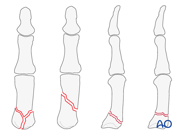
This approach is indicated for extraarticular fractures of the first metacarpal, and for periarticular fractures of the first carpo-metacarpal joint.
It is also a good choice for first carpo-metacarpal joint arthrodesis.
2. Surgical anatomy
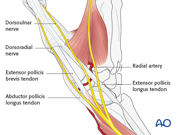
In the subcutaneous tissue, the dorsoulnar and dorsoradial divisions of the dorsal sensory branch of the radial nerve must be protected. The surgical approach is made between the tendons of extensor pollicis longus (EPL) and extensor pollicis brevis (EPB).
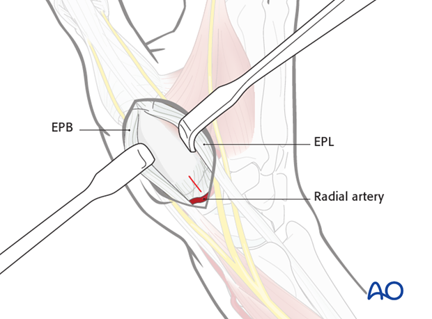
The radial artery crosses over the trapezium.
3. Skin incision
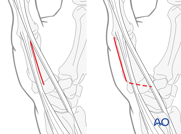
Make a straight skin incision over the interval between EPL and EPB throughout the length of the first metacarpal.
Alternatively, the incision can be made radial to the EPB. This incision can be extended proximally, curving in an ulnar direction.
Identify and protect the divisions of the dorsal sensory branches of the radial nerve, and the longitudinal veins.
4. Incision of the fascia
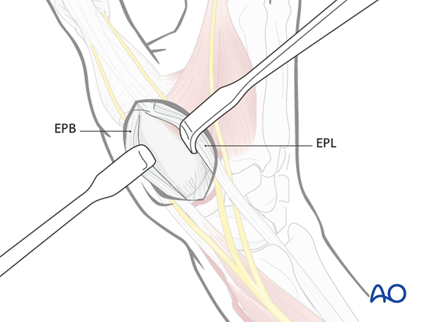
Incise the fascia between the EPL and EPB tendons and retract them to either side.
5. Alternative fascial incision
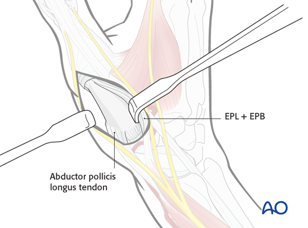
If the more radial skin incision has been chosen, incise the fascia on the radial side of EPB, and retract both tendons in an ulnar direction.
6. Option: capsulotomy
In the case of intraarticular fractures, or arthrodesis, a capsulotomy of the first carpo-metacarpal joint is made.
7. Wound closure
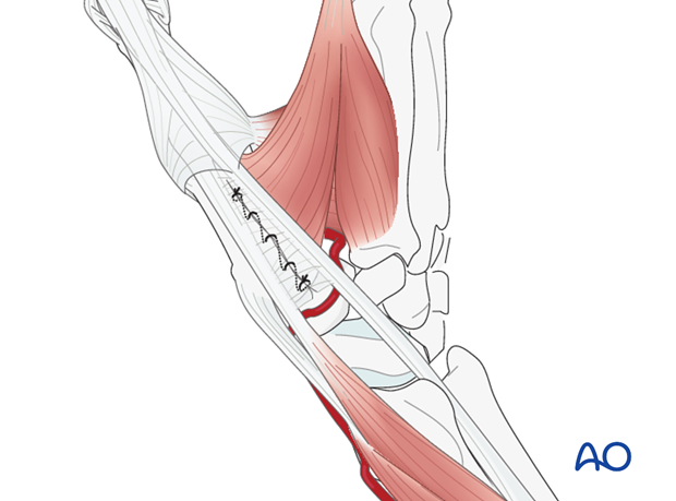
The fascia between the EPB and EPL tendons can be repaired either with a running suture or with multiple fine mattress sutures.
8. Pearl
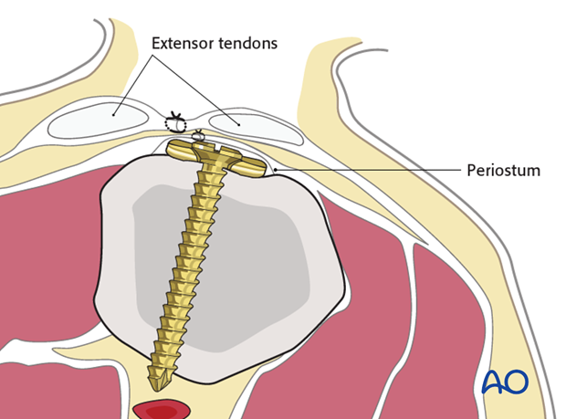
Lifting the periosteum and covering the implant with it, as far as possible, helps to minimize contact between extensor tendons and the implant. They will then glide over the periosteum, limiting additional injury.
