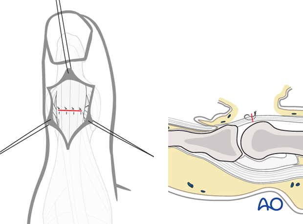
Dorsal approach to the IP joint of the thumb
1. Indications
This approach is indicated for
- Interphalangeal (IP) arthrodesis
- Intraarticular fractures of the interphalangeal joint of the thumb
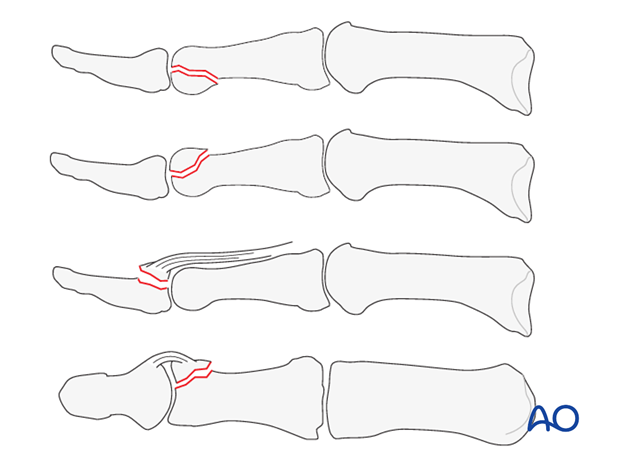
- Dorsal avulsion fractures of the base of the distal phalanx by the extensor pollicis longus (EPL) tendon
- Fracture avulsions of collateral ligament of the IP joint
2. Skin incision
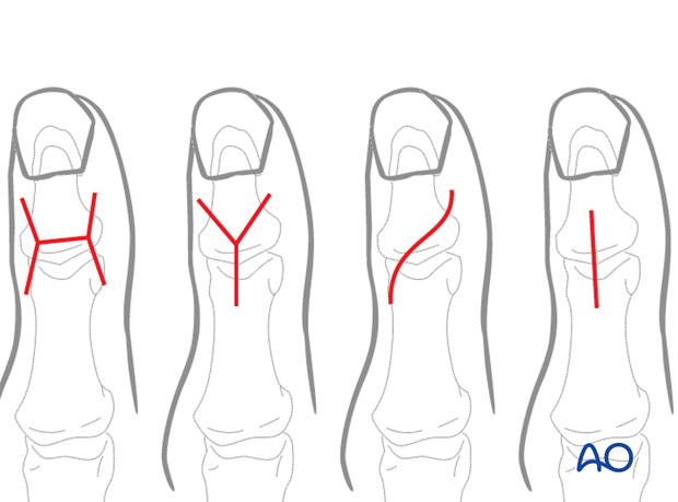
There are four common skin incisions: The H-shaped, the Y-shaped, the lazy S-shaped and midline longitudinal incisions.
The H-shaped incision is usually modified by diverging the sides slightly, to minimize vascular insult.
3. Transverse incision
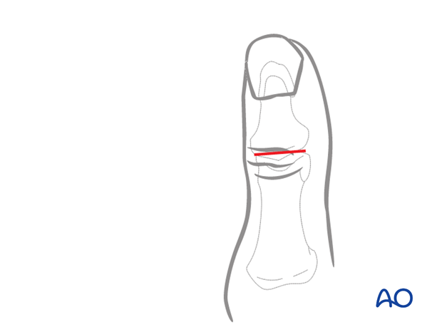
An alternative incision, designed to reduce potential soft-tissue trauma, is a simple transverse incision, which will usually afford enough exposure for osteosynthesis and joint surface preparation for arthrodesis.
Pearl
Note that the level of the joint line lies deep to the most distal of the dorsal extensor creases: it is a common mistake of the less experienced surgeon to place the transverse incision too proximal, thereby limiting access to the base of the distal phalanx.
4. Elevation of the skin flaps
Depending on the shape of the skin incision, flaps should be elevated and held with fine sutures to minimize soft-tissue trauma.
Tiny veins will appear, and should be coagulated with the bipolar forceps, as necessary.
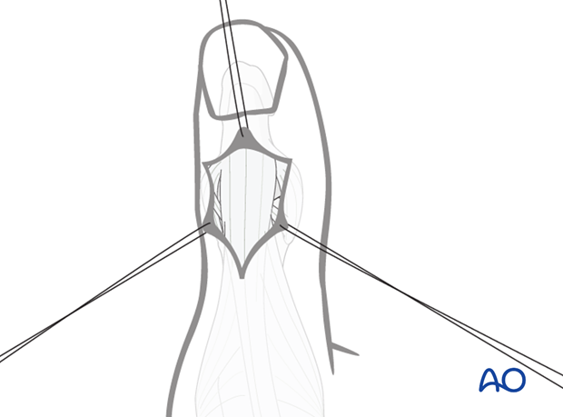
5. Division of the extensor tendon
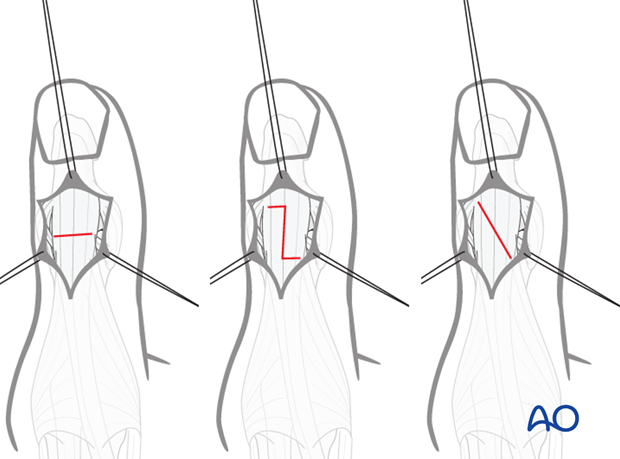
Divide the distal tendon of the EPL using either a transverse tenotomy, a step cut, or a long oblique cut.
The step cut and the oblique cut facilitate subsequent repair.
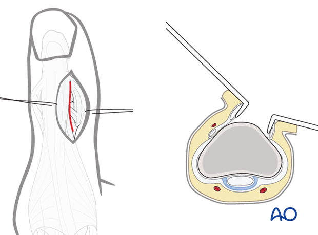
Alternative: retraction of tendon
A straight dorsoradial, or dorsoulnar, skin incision will allow retraction of the distal tendon of EPL without having to divide it.
This alternative only allows access to one condyle, but never to both.
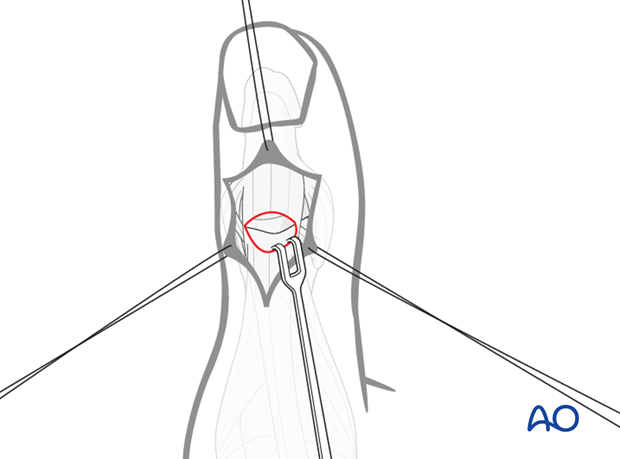
6. Exposure of the joint
Retract the EPL tendon proximally in order to expose the interphalangeal joint.
7. Wound closure
Repair the tenotomy using fine interrupted mattress sutures.
