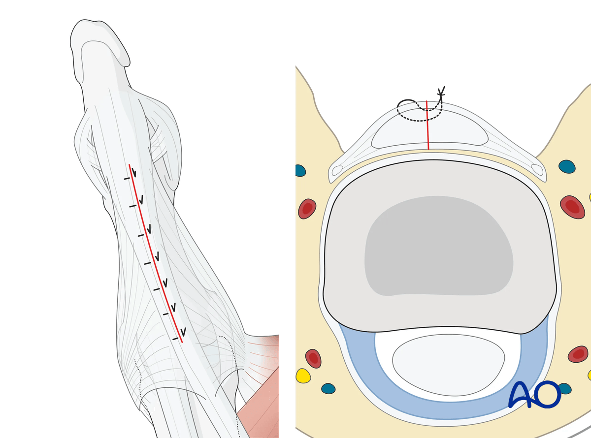
Dorsal approach to the proximal phalanx of the thumb
1. Introduction
The dorsal approach to the 1st proximal phalanx offers access to the extensor tendon and the dorsal aspect of the proximal phalangeal shaft. If extended, the metacarpophalangeal (MCP) and interphalangeal (IP) joints may be exposed.
2. Surgical anatomy
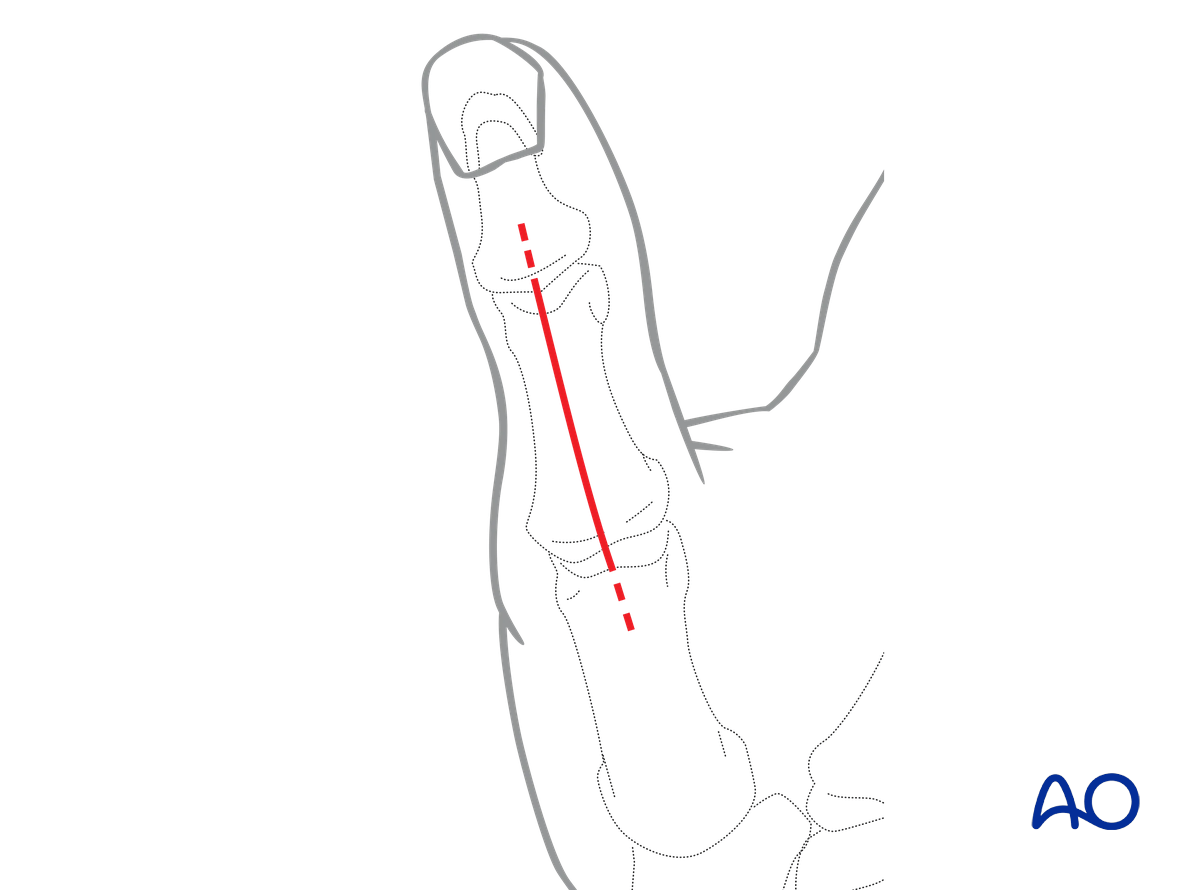
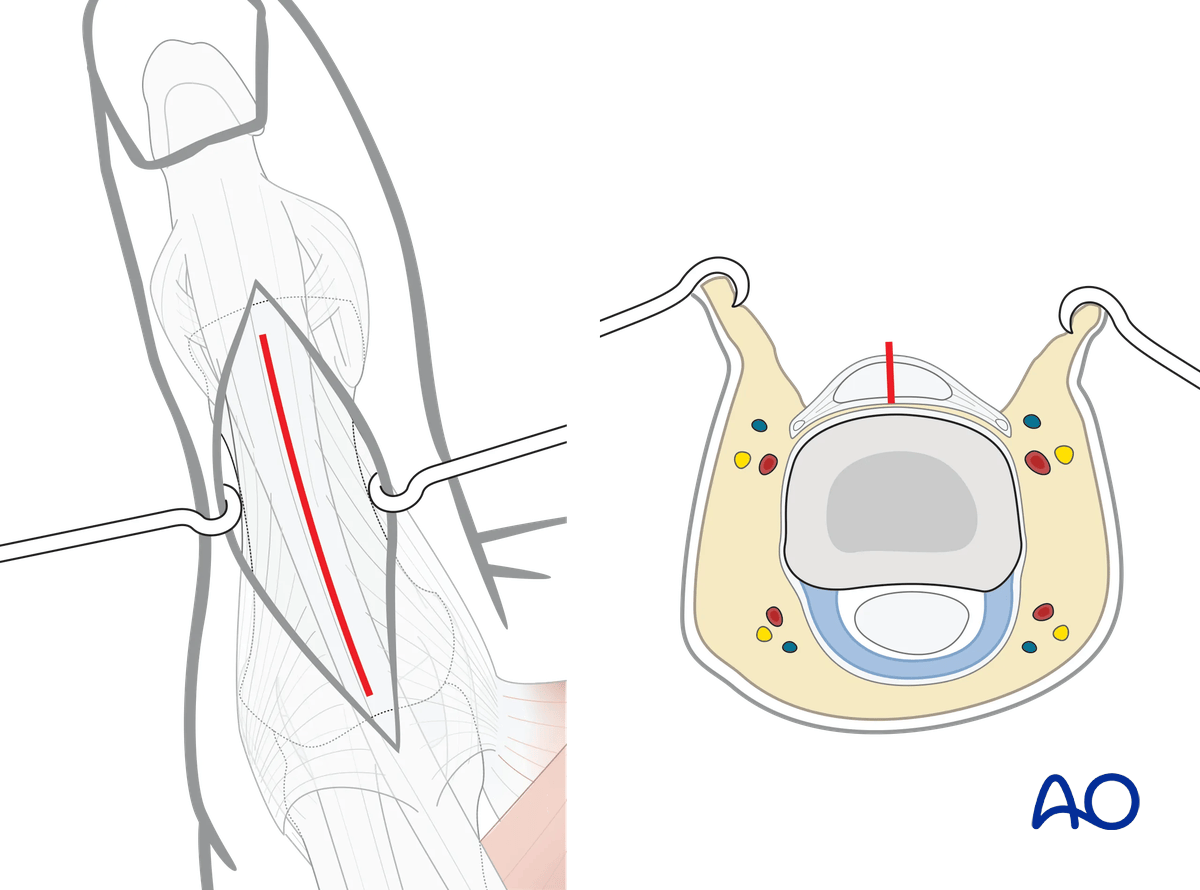
Full thickness skin flaps including the dorsal nerve branches are raised on both sides exposing the extensor mechanism.
If proximal extension over the MCP joint is planned, the position of the underlying nerve branches should be considered.
3. Skin incision
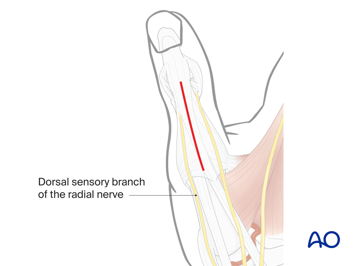
Perform a straight skin incision, starting at the MCP joint and ending at the proximal IP joint. Depending on the fracture morphology, the incision may be shorter.
With this incision, vascularity and venous drainage are well preserved. Early postoperative motion will prevent scarring between the skin, tendon, and bone.
The disadvantage of this incision is that any skin and tendon scarring will be in the same line.
Neural structures are well within the full thickness skin flaps.
4. Deep dissection
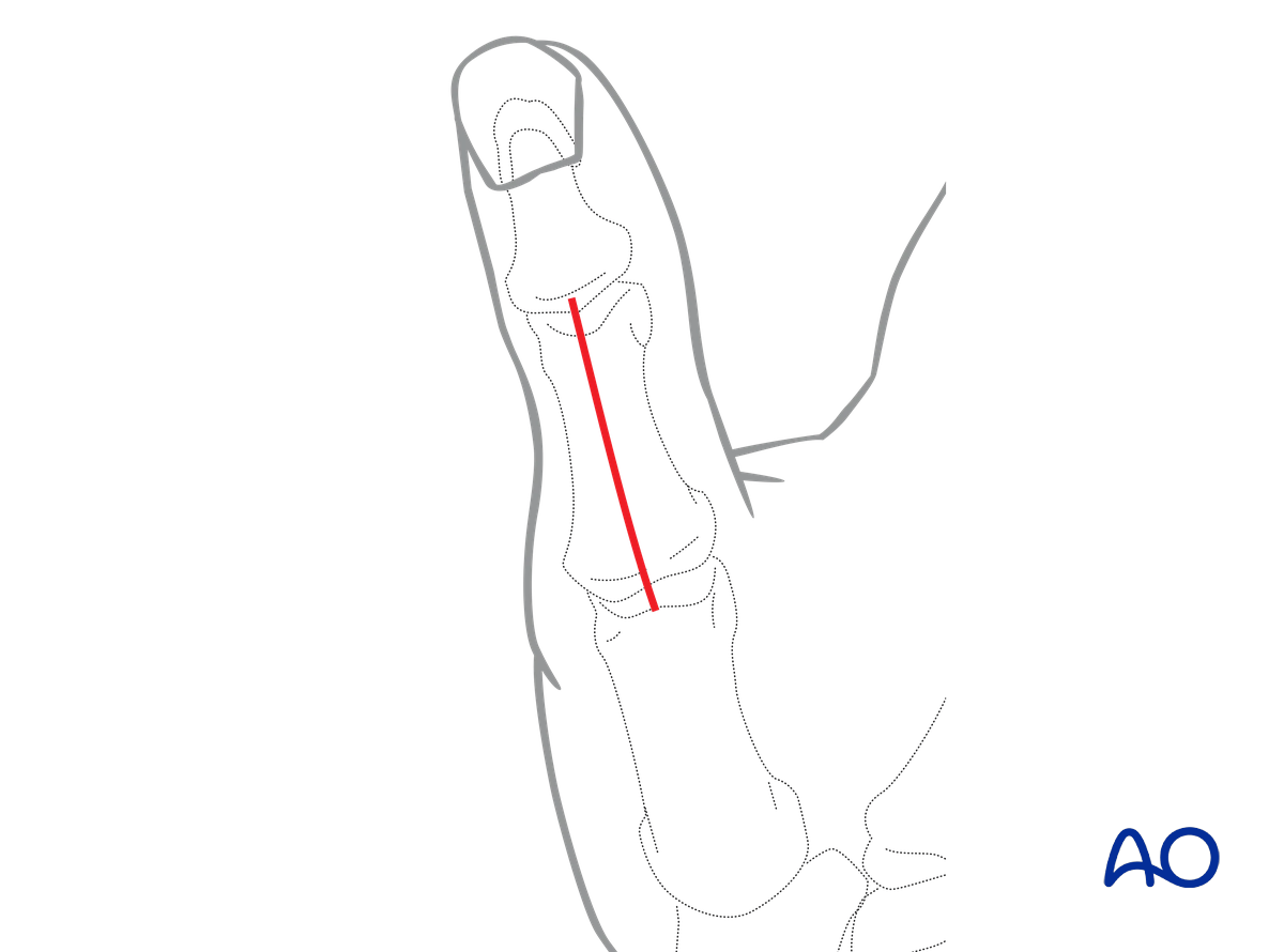
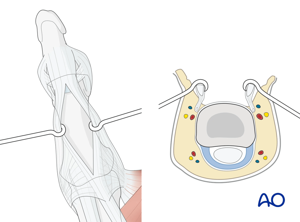
Split the extensor tendon apparatus along the midline longitudinal fibers of the tendon.
Avoid incising too distally, which could result in injury to the extensor tendon insertion at the base of the distal phalanx.
Fracture exposure
Retract the tendon and periosteum to expose the fracture site. Try to preserve as much of the periosteum as possible, which should be elevated only adjacent to the fracture line.
5. Wound closure
The tendon incision must be repaired with monofilament sutures with an atraumatic needle before wound closure.
Cover the implant with the periosteum as far as possible; this helps minimize contact between the extensor tendons and the implant.