ORIF - Screw fixation
1. Diagnosis
Mechanism of the injury
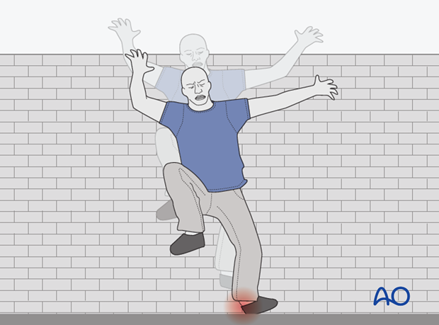
This fracture is often confused with an ankle sprain. The mechanism of this injury is an axial load. Diagnosis is often very difficult.
Imaging
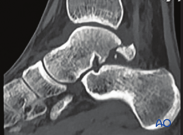
A plain x-ray may be diagnostic, but coronal CT scans are often necessary for a complete definition of the injury.
The presence of an os trigonum which is a developmental failure of the fusion of the posterior tubercle of the talus with its body may be very confusing in making a diagnosis. The aspect of the os trigonum facing the rest of the talus is corticated whereas a fresh fracture is not. If there is any doubt, a bone scan or an MRI would be definitive.
The x-ray shows a posterior process fracture. Case by courtesy of Dr. Steven Steinlauf, Florida, USA.
2. Surgical indications
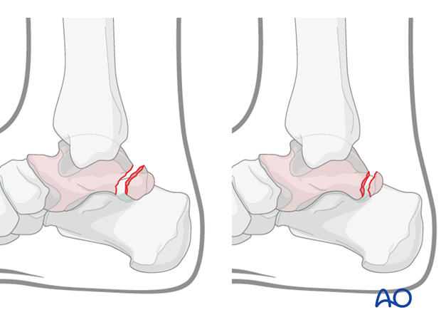
Fractures of the posterior process of the talus may involve the subtalar joint, if they are large.
Because this is an intraarticular fracture, if displaced, anatomic reduction and stable fixation has to be restored to prevent the development of posttraumatic arthritis. If the fracture is comminuted and reduction and fixation is not possible, then one should consider resection. Similarly, if a symptomatic nonunion develops, the fragment should be resected. If this piece is large, screw fixation may be necessary.
If the fracture is completely undisplaced, then immobilization and non-weightbearing is indicated.
3. Approaches
The posterolateral or posteromedial approaches allow visualization of this fragment.
4. Reduction and preliminary fixation
Reduction
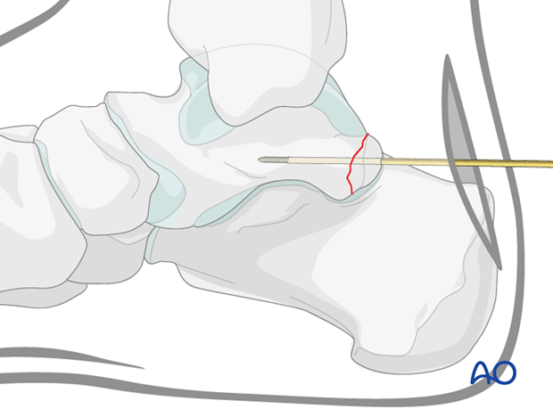
Because this fragment is frequently very small, a K-wire may be used as a joystick to help with the reduction which is carried out with the help of an image intensifier.
Preliminary fixation
The fragment will often not accept more than a single screw. Therefore it is best to use a threaded K-wire guide of a corresponding cannulated screw for provisional fixation. This threaded K-wire is then introduced into the middle of the fragment, once reduced, and advanced into the body of the talus. A cannulated lag screw can then be used for fixation.
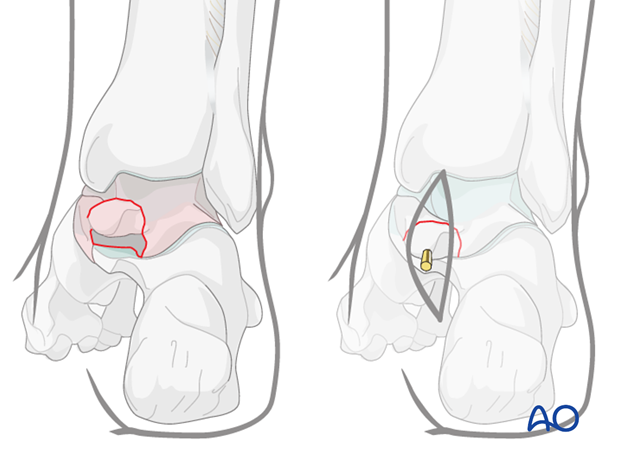
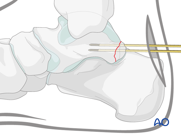
Preliminary fixation with eccentric K-wire
A second K-wire should be placed eccentrically, if possible, to provide rotational control of this small fragment of cortical bone. It will be removed when fixation is complete.
If possible, two screws would be optimal, but often not possible.
5. Screw fixation
Pearl: rule of thirds
This fracture is often quite small, or multifragmented.
If the piece is one large fragment, then screw fixation is preferred. If the piece is very small then resection may be a better option. K-wires alone may be used but present difficulty (migration) if used alone.
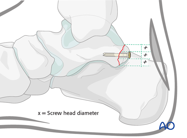
A nice rule to remember is the rule of thirds. If the size of the screw head is more than one third of the size of the fragment, then this small piece may comminute into many pieces. If the size of the screw head is less than a third of the fragment, then successful fixation may be achieved.
Drilling
With the central K-wire in place, drill the gliding hole with the corresponding drill bit, drilling only the fragment. Then drill the thread hole in the body of the talus.
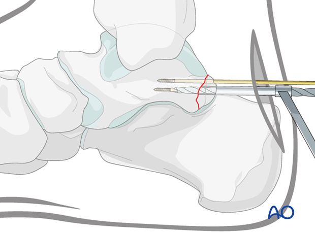
Screw insertion
Measure the depth for screw length.
A cannulated partially threaded screw will be used for fixation. Thus depending on the size of the fragment, one would use either the 2.4 mm, or the 3.5 mm screw and the corresponding drill bits.
Careful technique will ensure that the small posterior process does not fragment.
K-wires usually are removed so they do not migrate.
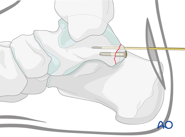
Second screw for rotational stability
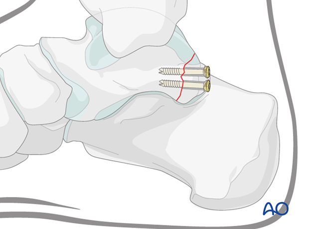
In order to secure rotational stability, it is necessary to insert a second screw, or if not possible, then a K-wire. Screws are preferable to a K-wire. Therefore try to insert a smaller screw to achieve rotational stability.
Use image intensification to make certain that reduction is anatomic and that the fixation has been appropriately inserted to maintain stability and articular congruity.
6. Aftertreatment
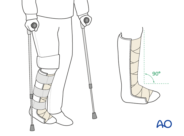
After surgery apply a posterior splint with the foot in neutral position. Early range of motion of the ankle and subtalar joint is advised.
Weight bearing should be restricted for 6 weeks with follow-up occurring at 2 and 6 weeks.
Radiography at 6 weeks should confirm healing. Once the fracture is united, progressive weight bearing and gait rehabilitation is started.
