- 1/3 – Decision making
- 2/3 – Casting
- 3/3 – Aftertreatment
1. Decision making
Radiology
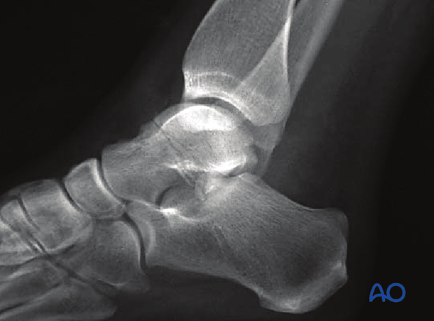
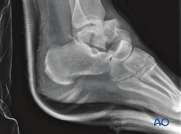
X-ray diagnosis is necessary using standard AP and lateral x-rays of the ankle and foot, with supplementary Canale views.
These x-rays are often difficult to interpret as there is more displacement or associated subluxation, or dislocation, of the ankle or hindfoot.
CT scans are often necessary.

If the talar neck fracture is undisplaced and all joint surfaces are in perfect alignment, then nonoperative treatment is a rational choice.
The patient must understand the importance of non-weightbearing compliance.

However, if this fracture is part of the spectrum of talar neck injuries which presents with displacement, requiring reduction, then there may be more decisions to be made about investigation and forms of treatment.
Plain x-rays may be all that is required if the fracture is undisplaced. This, however, is unusual as most talar neck fractures present with at least some displacement.
CT examination is very helpful whenever there is any question about displacement, or requirement for debridement of the subtalar joint. Along the spectrum of injury, increasing displacement presupposes that there is more subtalar and tibiotalar osteochondral injury. This often requires surgical approaches for debridement and fixation of these fracture types.

2. Casting
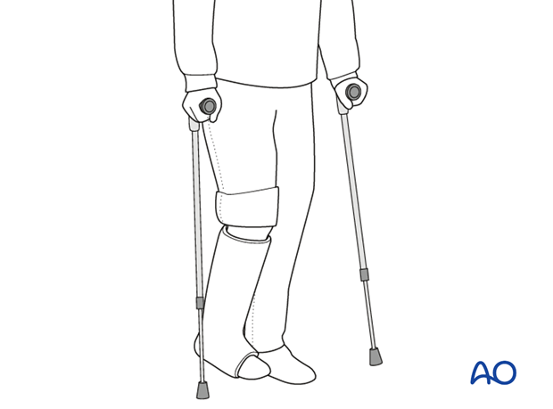
Undisplaced fractures are treated for 6-8 weeks in a non-weightbearing below-the-knee plaster cast. Patient compliance is mandatory and if the fracture displaces it results in surgery.
Avascular necrosis is unlikely with an incidence of 0-10%.

Teaching video
AO teaching video: Application of a Lower Leg Circular Cast
3. Aftertreatment
Non-weight bearing with a below-knee cast is continued for 6-8 weeks. Follow-up visits with radiography should occur at 2 and 6 weeks.
Once out of plaster, mobilization is started. Weight bearing is withheld until a full range of motion is achieved. In undisplaced fractures, full range of motion is usually achieved rapidly.
Gait training is initiated with physiotherapy, as required, at 6-8 weeks.


