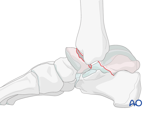
Neck, displaced
Decision making
If the fracture is displaced, then it is often associated with other hindfoot injuries. This will lead to more decisions about further investigation and other forms of treatment.
Plain x-rays may be all that is required if the fracture is undisplaced. This, however, is unusual as most talar neck fractures present with at least some displacement.
CT examination is very helpful whenever there is any question about displacement, or requirement for debridement of the subtalar joint. Along the spectrum of injury, increasing displacement presupposes that there is more subtalar and tibiotalar osteochondral injury. This often requires surgical approaches for debridement and fixation of these fracture types.
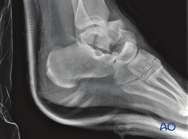
If the talar neck fracture is undisplaced and all joint surfaces are in perfect alignment, then nonoperative treatment is a rational choice.
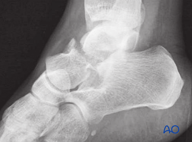
Fracture occurrence
Talar neck fractures occur with axial load and a foot with muscles in tension holding the foot in a rigid position. Commonly, motor vehicle crashes cause the foot to be axially loaded with the foot plate of the car impacting a dorsiflexed foot on the brake pedal. Associated soft-tissue injury can occur, especially with increasing severity. Open talar neck injuries or other foot and ankle pathology often are part of a spectrum of injury.

Radiology
X-ray diagnosis is necessary using standard AP and lateral x-rays of the ankle and foot, with supplementary Canale views.
These x-rays are often difficult to interpret as there is more displacement or associated subluxation, or dislocation, of the ankle or hindfoot.
CT scans are often necessary.
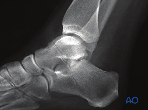
Anatomy
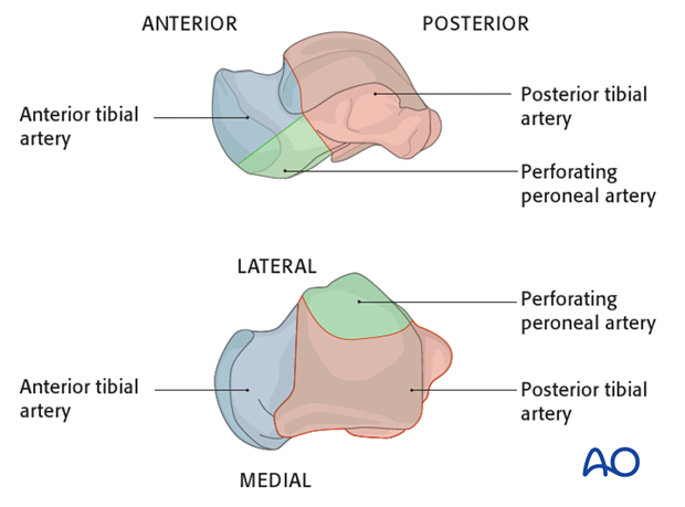
The blood supply to the body of the talus is complex. Most of the blood supply to the body is through the deltoid branches of the posterior tibial artery. The dorsalis pedis and peroneal artery supply the neck and the lateral third. Fracture dislocations can easily compromise the blood supply to the body of the talus and lead to avascular necrosis.
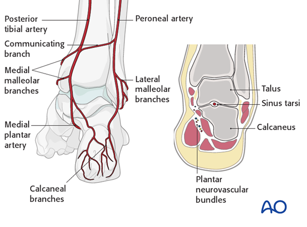
Note the deltoid branches of the posterior tibial artery and the artery of the tarsal canal. A posteromedial approach to the body of the talus would destroy its blood supply. Therefore, if the body has to be exposed, one utilizes an osteotomy of the medial malleolus.
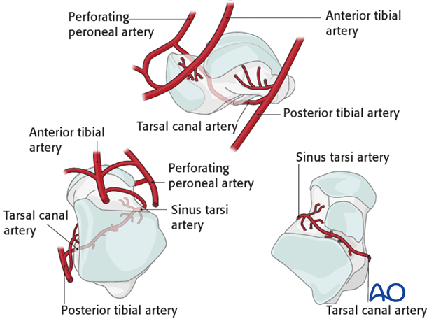
The deltoid branches are important to supply blood to the medial talar neck and talar body. Branches from the dorsalis pedis supply the talar head and most of the dorsal talar neck. The artery of the tarsal canal coming from branches off of the posterior tibial artery supply most of the talar body.
The peroneal artery has the least contribution laterally.
Classification
The Hawkins classification is based upon blood supply to the talus.
In type 1 injuries, the medial blood supply is still assured.
In type 2 injuries, the medial blood supply may be preserved.
In type 3 injuries, all medial blood supply to the body is disrupted.
The type 4 injury, in which the body of the talus is completely dislocated and the head fragment displaced, has the worst prognosis because of avascular necrosis of the body and often of the head fragment.
Type I
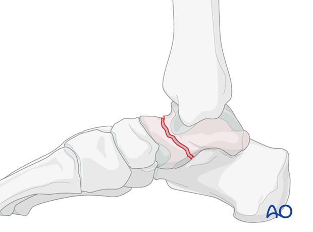
Nondisplaced fracture of the talar neck
Type II
Displaced fracture of the talar neck with subluxation or dislocation of the subtalar joint
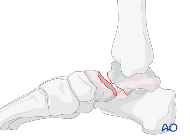
Type III
Displaced fracture of the talar neck with dislocation or subluxation of the talar body from both the tibiotalar and subtalar joints
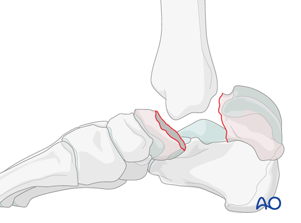
Type IV
Displaced fracture of the talar neck with dislocation or subluxation of the talonavicular, tibiotalar, and subtalar joints
