Osteonecrosis
Introduction
When a bone fractures and its blood supply is damaged then osteonecrosis can occur. Certain bones such as the talus, femoral head, scaphoid and proximal humerus are susceptible.
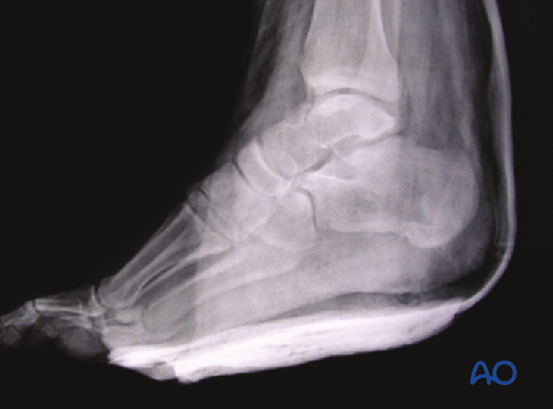
This x-ray shows a Hawkins II talar neck fracture, presurgery.

Risk factors of osteonecrosis
With increasing displacement and comminution, osteonecrosis becomes more prevalent. Hawkins type I incidence is less than 10% while Hawkins type IV incidence nears 100%.
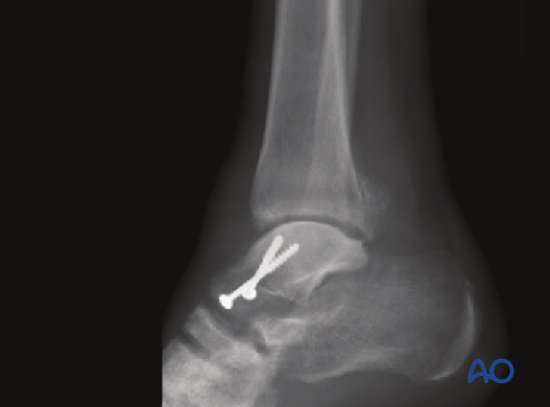
This x-ray shows the same patient, three months postsurgery with avascular necrosis of the talar body.

Diagnosis of osteonecrosis
When a patient presents at 6-12 weeks for follow up with a bone that is at risk for osteonecrosis, then the radiologic sign (Hawkin’s sign) is present. This means that subchondral lucency appears immediately beneath the joint surface signifying the bone is alive. But if this lucent line does not appear then avascular necrosis may be present. If there is chondral collapse with the revascularization of the avascular bone then osteoarthritis will develop.
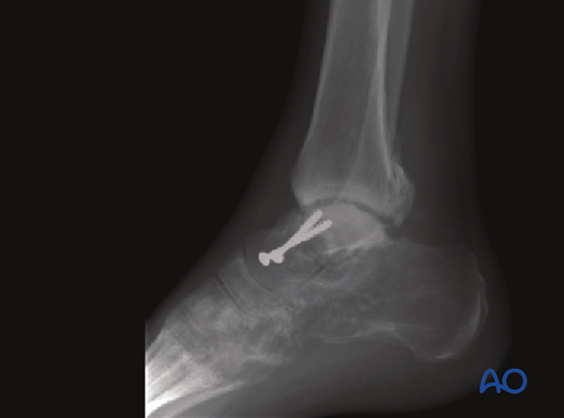
This x-ray shows at 5 months avascular necrosis of the talus with early osteoarthritis in both the ankle and subtalar joint.

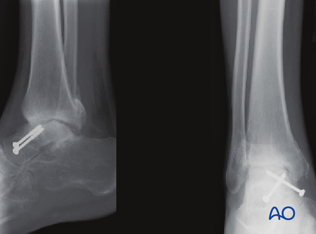
This x-ray shows at 8 months irreversible osteoarthritis of both ankle and subtalar joint.

Long term treatment
Osteonecrosis is rarely preventable and usually progressive. Its treatment is benign neglect and nonoperative including bracing, anti-inflammatory medication and supportive care. Eventually some joints need to be fused to eliminate joint pain.


