Direct lateral approach to the talus
1. Introduction
The direct lateral approach is indicated to expose a simple fracture of the lateral process of the talus or to debride the subtalar joint.
Although very similar to the middle section of the anterolateral approach, this approach is positioned a little more plantar to access the lateral process of the talus. Fluoroscopy is useful when planning the incision.
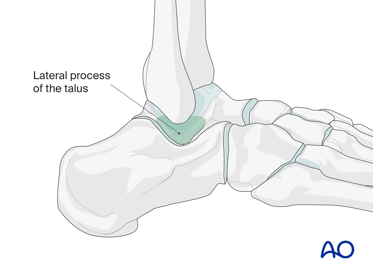
It utilizes a window (marked in green) to approach fractures of the lateral process of the talus (eg, “snowboarder’s fracture”). Arthrodesis or arthroscopy of the posterior talocalcaneal joint can be performed with this direct lateral approach.
2. Anatomy
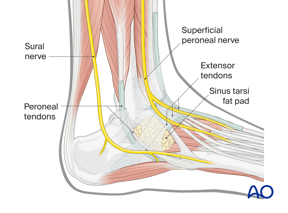
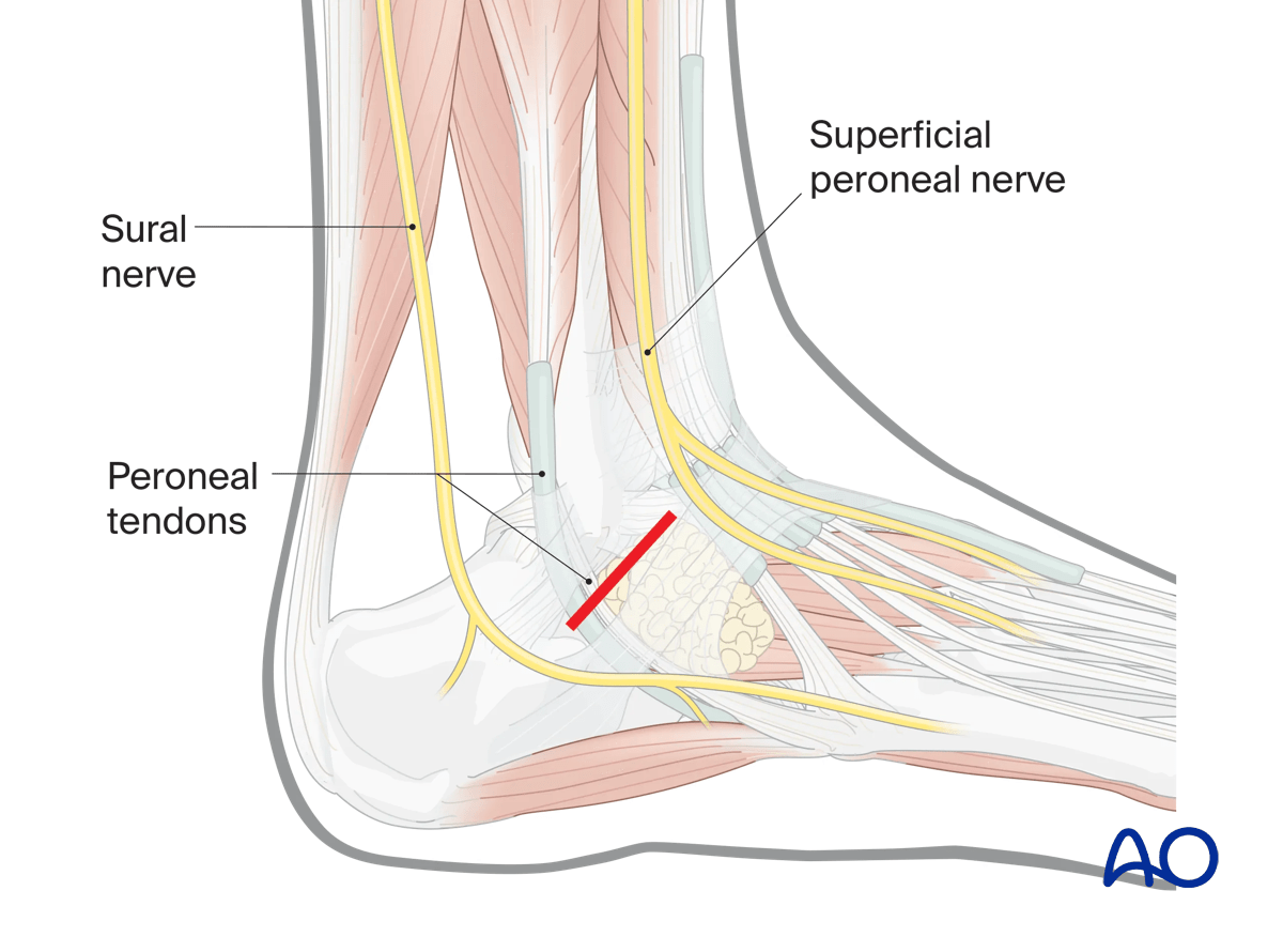
There are few important structures in this area. Note the peroneal tendons posteriorly and laterally, and the extensor tendons more anteriorly and dorsally.
Between these two tendons is a fat pad which leads one to the subtalar joint.
3. Incision
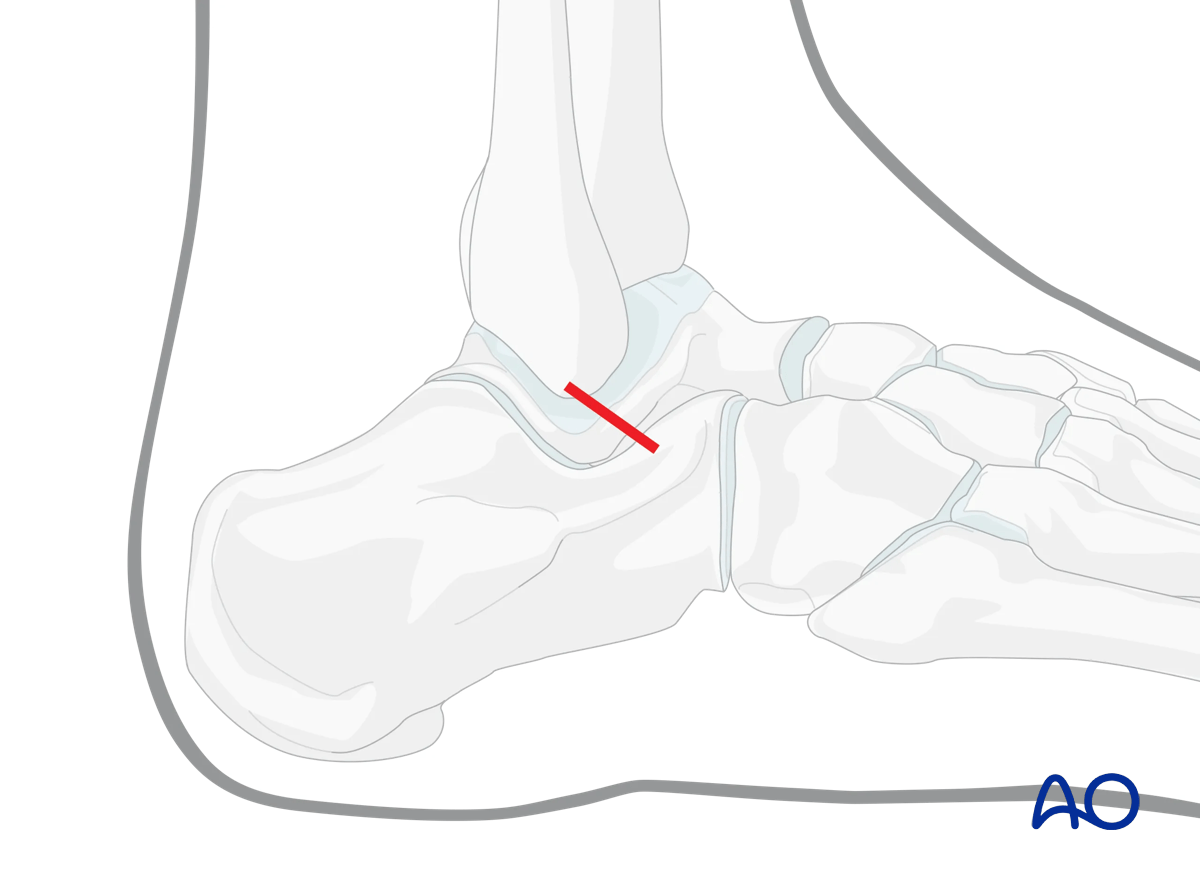
This incision may be performed either longitudinally parallel to the tendons or transversely.
Here, a longitudinal incision is shown. An image intensifier with a lateral projection will facilitate precise incision placement.
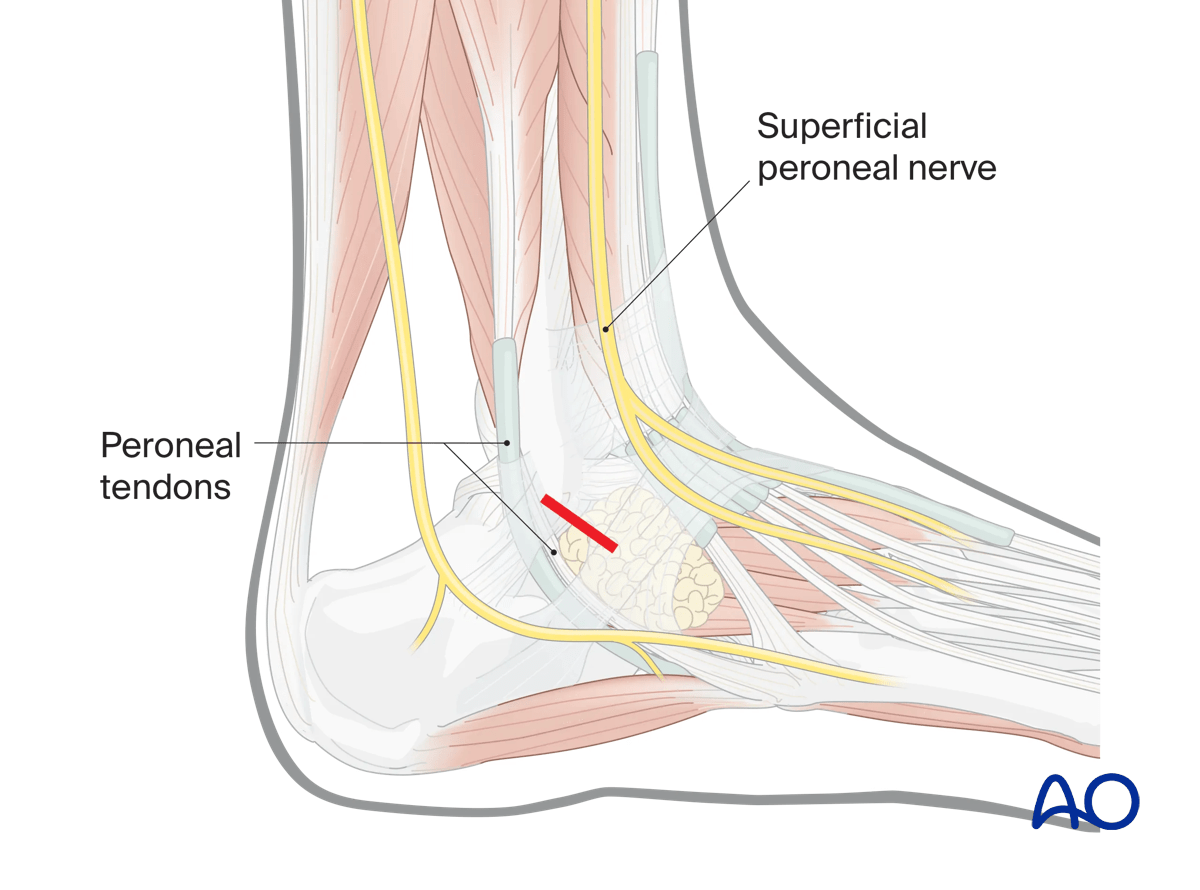
A transversely oriented incision is illustrated here.
Note that it is limited posteriorly by the sural nerve and anteriorly by the superficial peroneal nerve. It is important to localize the length and position of the incision properly.
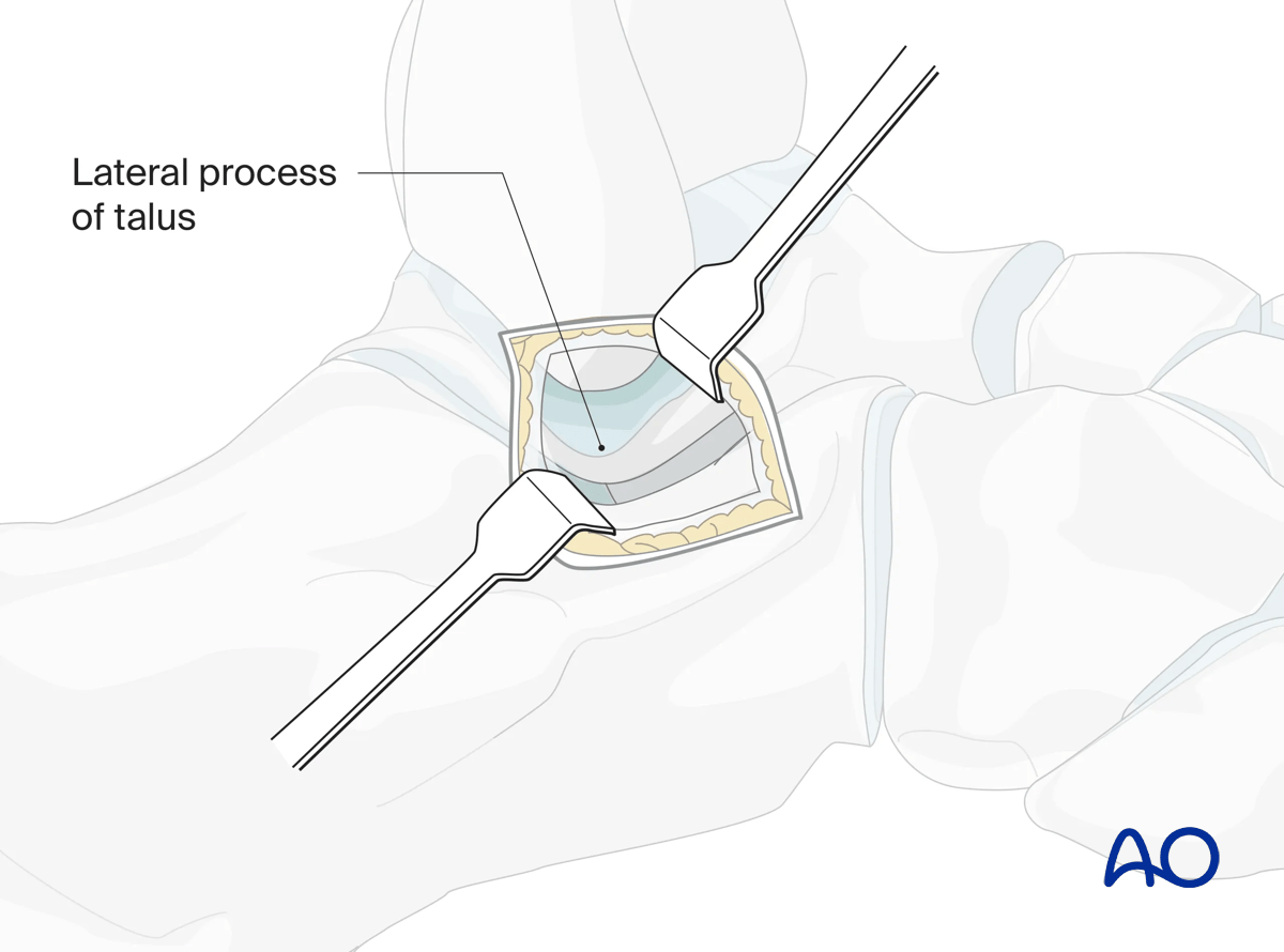
Once through deep fascia, an area of fat is found. Incise the fat pad to reveal the subtalar capsule, the lateral talar process.
4. Wound closure
This approach is closed in layers.


