Anterolateral approach to the talus
1. Indications
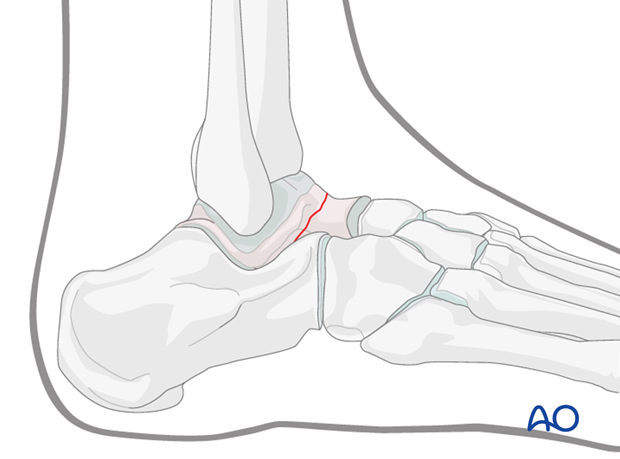
The anterolateral approach is usually employed in combination with the anteromedial approach to gain access to any displaced talar neck fracture.
However, for certain fractures such as minimally displaced fractures or very simple neck fractures, it can be used as the sole approach.
2. Anatomy
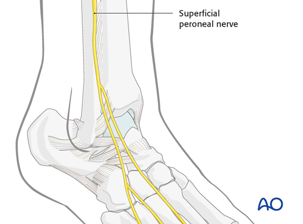
Around the ankle and hindfoot, full-thickness incisions without undermining are imperative. To avoid cutting the branches of the superficial peroneal nerve, most of the incisions must be made in a longitudinal direction.
The superficial peroneal nerve lies in the incision or very near and must be avoided and protected.
3. Incision
Skin incision
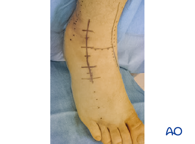
The incisions here are based on the deep talar anatomy which lies underneath the extensor digitorum brevis.
The incision is based on the fourth metatarsal and lines up with this bone.
Deep dissection
The muscle of extensor digitorum brevis is bulky but once this muscle is split, …
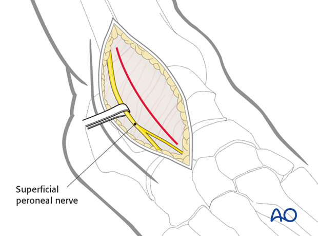
… one gains access to the lateral talus and subtalar joint.
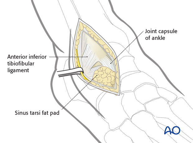
4. Exposure of the anterolateral talar neck
Once the extensor digitorum brevis is split longitudinally and retracted, one exposes the lateral aspect of the talar neck.
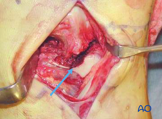
Most talar neck fractures result from a medially directed force. Therefore, the lateral side of the neck comes under tension and the medial side under compression. The fractures on the lateral side are simple, and on the medial side multifragmentary. Exposure from the medial side alone runs the risk of shortening the neck and malreducing the fracture. To ensure an anatomic reduction, fractures of the neck must be exposed from the lateral and medial side.
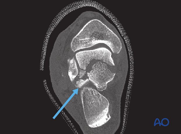
The arrow on the image indicates a simple fracture of the talar neck.
5. Debridement of subtalar joint
Debridement of the subtalar joint is imperative to facilitate an excellent reduction.
Using both anterolateral and anteromedial approaches provides excellent access to the whole talar neck. The importance of this lies in getting a perfect reduction for definitive fixation.
Our CT image shows comminution of the inferior talus.
