Clinical and radiological examination of patients with scapular injuries
1. Clinical examination
Common associated injuries which must be excluded are the following:
- Shoulder dislocation and associated articular lesions such as labral tares and rotator cuff lesions
- Proximal humeral fractures
- nerve lesions or plexus palsies
- Vascular injuries
- Upper thorax trauma
2. Radiological examinations
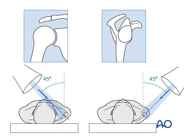
For the diagnosis of scapular injuries two X-ray projections at 90 degrees to one another are routinely taken:
- 45 degree antero-lateral (trans glenoid view or epsilon view
- 45 degrees antero-medial (trans scapular view)
The trans glenoid or epsilon view is taken with the beam at 45 degrees to the sagittal plane. The anode is positioned anteriorly and the cathode posteriorly in line with the orientation of the scapula. This anterolateral orientation of the C-arm will produce a trans glenoid projection and an AP of the glenoid neck and body of the scapula.
In order to get a trans scapular view which is necessary to evaluate any cranial or caudal displacement of either the joint block or fragments of the body of the scapula, we position the C-arm in such a way that the beam is at right angles to the scapula. The anode is usually positioned lateral and posterior, and the cathode medial and anterior. The beam is directed medially going perpendicular to the scapula.

In order to get detailed information a CT of the scapula is mandatory. If reconstructions are available these are to be ordered and sometimes a 3D reconstruction is useful.


