Arthroscopic approach to the knee
1. Principles
The arthroscopic approach is only recommended in minimally, or nondisplaced, fractures in young patients. Advanced experience in arthroscopic surgery is essential.
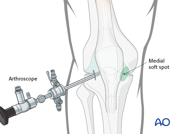
2. Anterolateral port
Identify the lateral soft spot adjacent to the patellar tendon.

Make a 5 mm stab incision and insert the arthroscopic shaft carefully, so as not to damage the intraarticular cartilage surface.
Next, remove the trocar and insert the arthroscope.
Irrigate the knee joint cavity until clear visibility is achieved.
Continuous positive pressure pump irrigation is essential, in order to prevent continuous bleeding, maintaining a pressure of 50-60 mmHg. High flow rates are required and adrenaline may be added to the saline for vasoconstriction.
The arthroscopic approach always requires that a full intraarticular examination of the knee joint is done, in order to avoid missing meniscal tears and ligament injuries, as well as osteochondral flake fractures.

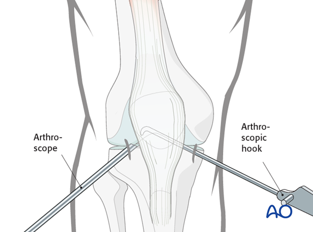
3. Anteromedial port
Identify the medial soft spot adjacent to the patellar tendon and palpate under arthroscopic control.

Make a 5 mm stab incision and insert the arthroscopic hook.
A small reduction tool may go in through the medial portal and the tibial spine avulsion is reduced.

A small stab incision is made if needed to allow for the introduction of a lag screw from medial peripatellar in the proximal tibia in a retrograde fashion.

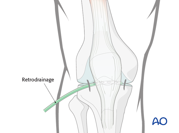
4. Wound closure
After removal of the arthroscope, insert a suction drain through the arthroscopic shaft.
Close the skin incisions with sutures in a routine manner.


