Limited open reduction and preliminary fixation of lesser tuberosity and humeral head
1. Principles
Lesser tuberosity reduction
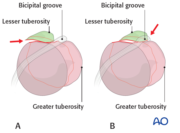
Proper reduction of the lesser tuberosity is difficult. Its position is hard to visualize with intraoperative image intensification.
Medial displacement of the lesser tuberosity (A) produces an intraarticular anterior step which can compromise internal rotation.
Lateral displacement of the lesser tuberosity (B) obstructs the bicipital groove and may compromise the biceps tendon. If possible, correct reconstruction of the bicipital groove is desirable to allow sliding of the tendon. An alternative would be tenodesis of the long head of the biceps.
2. Reduction and suture fixation of lesser tuberosity
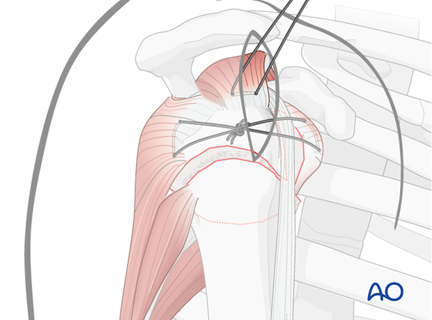
Sutures in the subscapularis tendon insertion aid both reduction and fixation of the lesser tuberosity. Once reduced, lesser tuberosity sutures are tied to a similar suture in the infraspinatus tendon for provisional fixation. Ultimately, these sutures contribute significantly to primary stability of the lesser tuberosity.
Placement of rotator cuff sutures
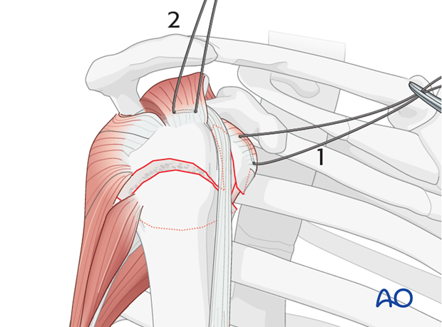
Subscapularis and supraspinatus tendon
Begin by inserting sutures into the subscapularis tendon (1) and the supraspinatus tendon (2). Place these sutures just superficial to the tendon’s bony insertions. These provide anchors for reduction, and temporary fixation of the greater and lesser tuberosities.
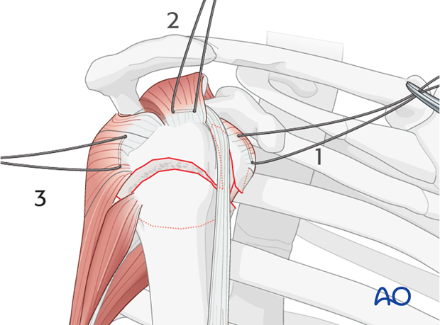
Infraspinatus tendon
Next, place a suture into the infraspinatus tendon insertion (3). This can be demanding, and may be easier with traction on the previously placed sutures, with properly placed retractors, and/or repositioning the arm.
Use of stay sutures
Anterior traction on the supraspinatus tendon helps expose the greater tuberosity and infraspinatus tendon.

Insert a preliminary traction suture into the visible part of the posterior rotator cuff …
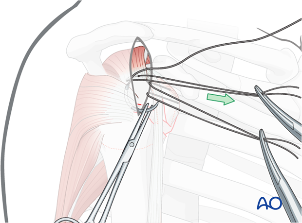
… and pull it anteriorly. This will expose the proper location for a suture in the infraspinatus tendon insertion. Then the initial traction suture is removed.
Pearl: A stout sharp needle facilitates placing a suture through the tendon insertion.
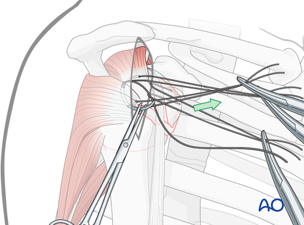
Reduction of lesser tuberosity
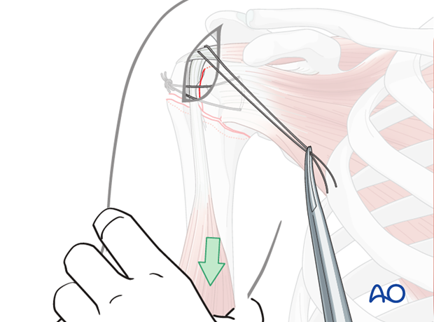
Direct reduction of the lesser tuberosity fragment is performed by pulling the sutures or, …
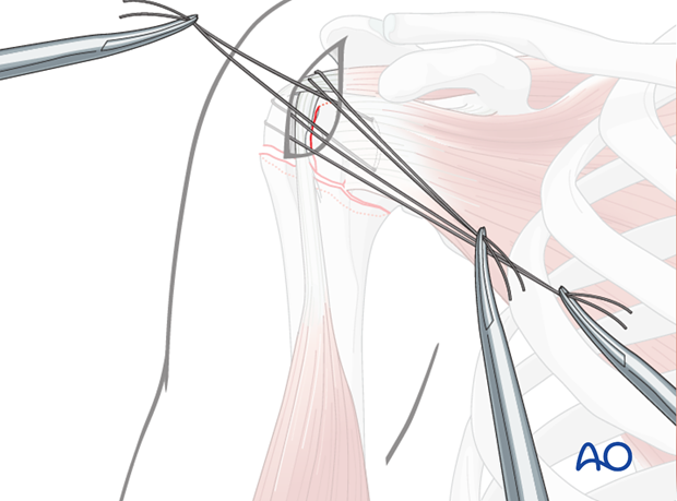
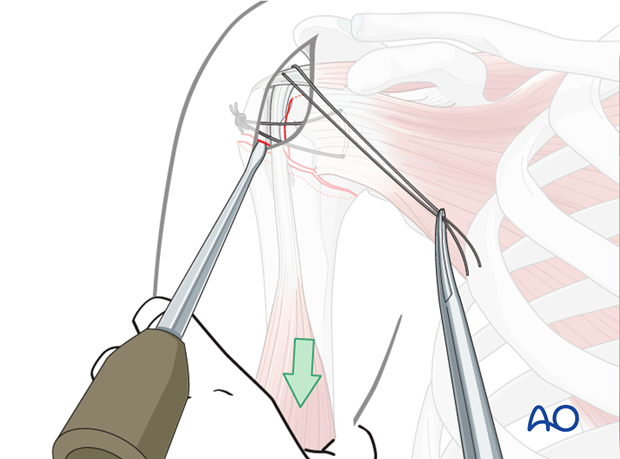
… with instruments (eg, elevator) applied either through the incision (as illustrated) or through a separate stab incision.
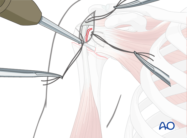
Preliminary fixation of lesser tuberosity
Tighten and tie the transverse sutures to preliminarily fix the lesser tuberosity fragment. Thereby, the 3-part fracture is converted into a 2-part situation.

3. Reduction and preliminary fixation of the head fragment
Reduction with traction
Distal traction, perhaps augmented with increased angulation, will help to reduce the fracture.
Use of an elevator
Sometimes, the incision allows insertion of an elevator to disimpact the humeral head, or to help to correct inclination/torsion and to restore a normal relationship of the medial fracture surface. The proximal fragment should be reduced anatomically to the shaft.
The actual process of reduction is done with image intensifier control.
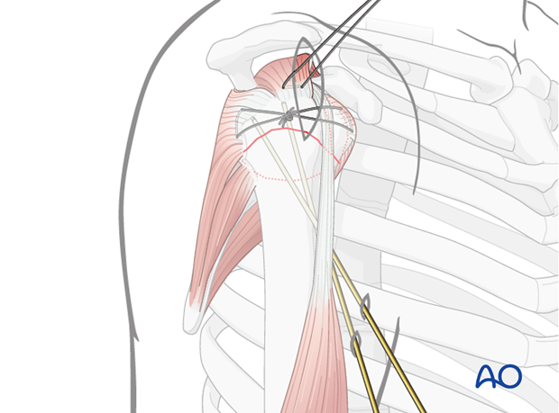
Preliminary fixation
After the tuberosity and humeral head have been reduced and stabilized with sutures, there may be no need for additional preliminary fixation, but it might be advantageous to use additional K-wires to secure the position of the humeral head relative to the humeral shaft. This is illustrated with 2 retrograde K-wires. Make sure to place them from anterior to avoid interference with the foreseen plate position.
4. Confirmation of reduction
Confirm the correct reduction in both AP and lateral views using image intensifier control.


