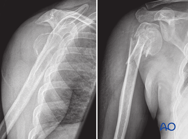
4-Part, marked displacement, fragmentary articular, varus malalignment
These fractures include a transcephalic (head-splitting) fracture line. This runs obliquely, somewhat parasagittally. A significant portion of the head remains attached to the greater tuberosity. A radiographic indication of a transcephalic fracture is a double profile of the convex subchondral bone of the humeral epiphysis. Optimal treatment is anatomical reduction and stable fixation. Arthroplasty may need to be considered in the elderly, osteoporotic patient.
This group of 4-part fractures is characterized by a marked displacement of the fragments and disruption of the periosteal sleeve. They are highly unstable. Due to the disrupted periosteal sleeve, especially on the medial side, the blood supply of the humeral head is severely affected. Prognostic factors of an ischemic humeral head are a) a fracture line located in the anatomic neck, b) a ruptured medial soft-tissue hinge, and c) a short posteromedial distal metaphyseal extension beyond the humeral head (less than 8 mm). Although these fractures do not necessarily pass through the articular surface, they are termed “articular fractures”; the articular surface is detached from both tuberosities and/or fractured itself. 4-part fractures according to Codman and Neer separate the proximal humeral epiphysis from both tuberosities and the metaphysis.

X-rays by courtesy of B Ockert, LMU Munich, Germany.


