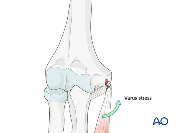
Repair of medial collateral ligament
1. Principle
The collateral ligaments of the elbow will heal at proper tension if the elbow remains concentrically reduced for 3 to 4 weeks. This is true even when the elbow has been dislocated for several months.
Some surgeons have reported the use of tendon grafts to treat acute, subacute, and chronically dislocated elbows. In the acute stage this is presumably to add additional stability.
It’s much easier to use strategies for maintaining elbow reduction (repair or replacement of all fractures, suture reattachment of avulsed collateral ligaments, and selective temporary mechanical maintenance of reduction (eg cross pins, external fixation, internal fixation)).
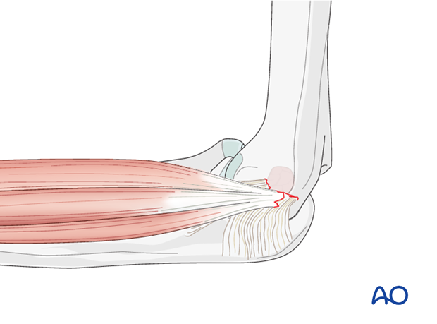
2. Preparation
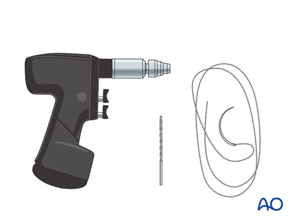
Instruments and implants for suture fixation
Sutures are generally used in cases of pure ligamentous avulsion without a bony fragment. This technique can also be used to fix a small bony fragment.
0 or 1 resorbable monofilament suture is generally recommended.
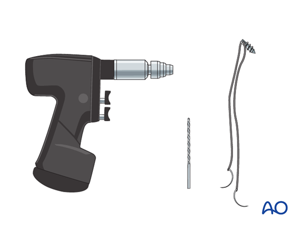
Instruments and implants for anchor fixation
It is recommended that one or two anchors are used. While one anchor may be sufficient, it is recommended that two are used where possible.
The potential disadvantage of anchor fixation is the prominence of the anchor-suture combination. The suture is not resorbable and if prominent and/or painful may require subsequent removal.
Anesthesia and positioning
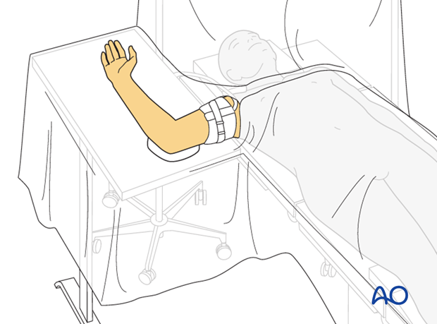
General anesthesia is recommended and a sterile tourniquet should be available.
The patient is placed supine with the arm draped up to the shoulder.
3. Exposure
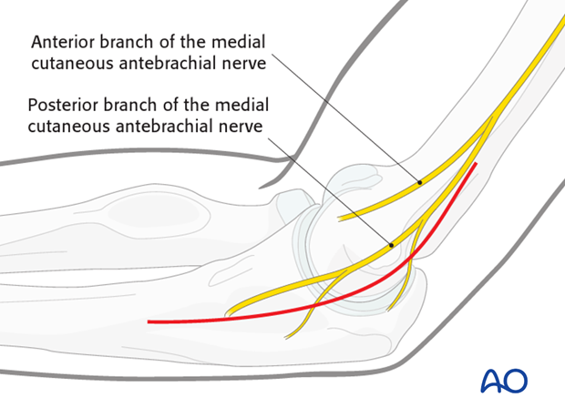
The skin incision can either be posterior with a medial skin flap or direct medial, taking care to protect branches of the medial antebrachial cutaneous nerve.
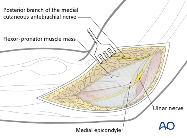
Ulnar nerve
For safe, facile access to the medial epicondyle the ulnar nerve should be released and transposed anteriorly or posteriorly while the repair is completed. Take care to use oscillating drills to protect the nerve. At the end of the surgery the nerve can be left anteriorly transposed in the subcutaneous tissues or replaced back in the groove if the surgeon prefers, and it does not subluxate with elbow flexion.
4. Repair
Suture repair
A figure-of-eight suture is inserted into the ligament and avulsed bone.
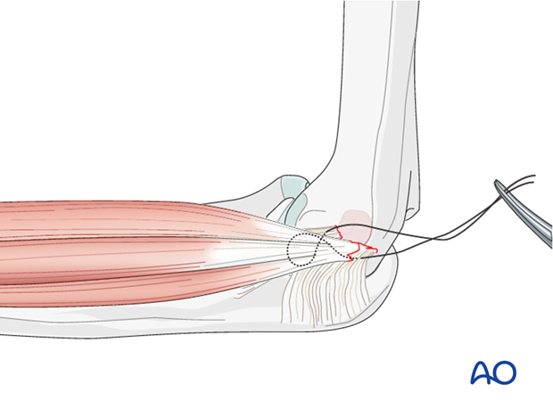
Two drill holes are made at the medial epicondylar defect (as shown in the illustration).
Take care to use oscillating drills to protect the nerve.
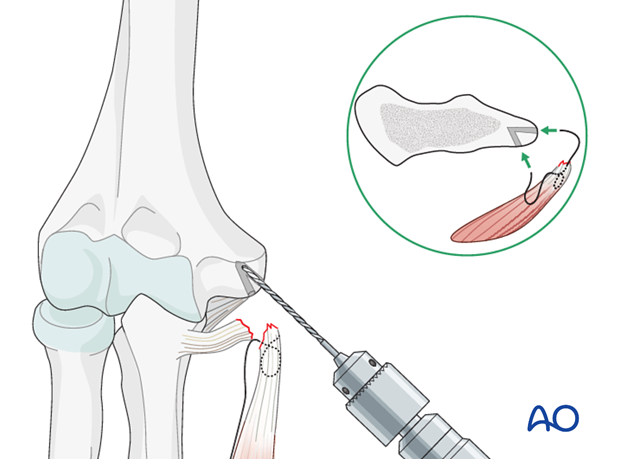
The needle of the suture is passed through these holes.
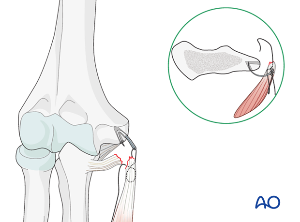
Pearl: The forearm is supinated and a varus stress is applied to the elbow to reduce tension in the flexor muscle mass prior to ligament reduction and reattachment (demonstrated here by the green arrow).
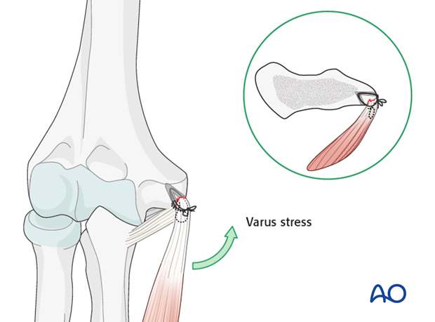
5. Anchor fixation
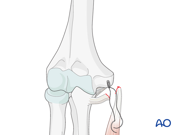
One (or two) drill holes are made in the medial epicondylar defect to accommodate the size of the anchors (this step is not necessary if self-drilling, self-tapping anchors are used).
Take care to use oscillating drills to protect the nerve.
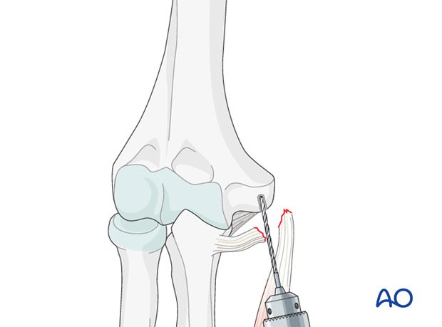
The anchor is inserted and fixed.
Pearl: The forearm is supinated, and a varus stress is applied to the elbow to reduce tension in the flexor muscle mass prior to ligament reduction and reattachment (demonstrated here by the green arrow).
The ligament and avulsed cartilage/bone are transfixed with the suture in a figure-of-eight pattern.
