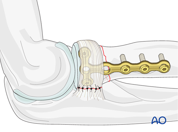
Radius, extraarticular, transverse - Compression with T-plate and screws
1. Preliminary remarks
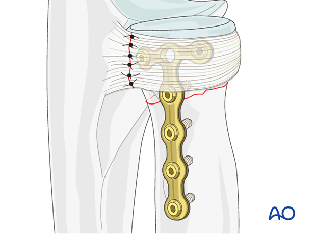
In transverse or short oblique fractures of the radial neck, fixation of the radial head to the diaphysis is achieved by plating.
2. Reduction and preliminary fixation
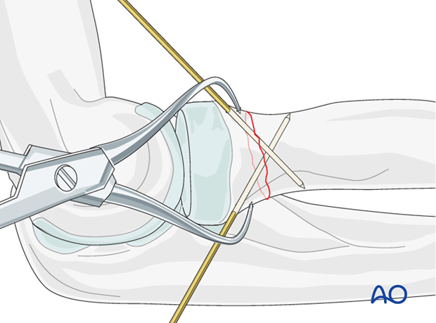
Expose the fracture ends with minimal soft tissue dissection off the bone.
Clean out the debris to allow perfect reduction.
Reduce the fracture with the help of small pointed reduction forceps and provisionally fix it with one or two 1.0 mm K-wires or reduction clamps.
Anticipate the plate position when placing the K-wires to avoid interference. In situations with an associated ulnar fracture, ulnar reduction must be perfect, otherwise radial neck anatomic reduction together with radio-humeral joint congruency will not be possible.
3. Plate positioning
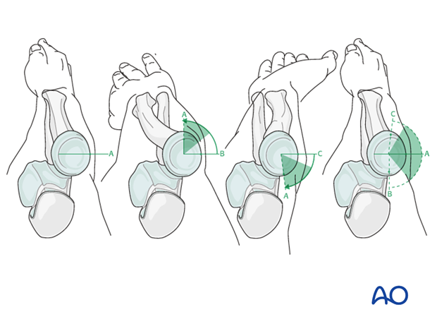
The radial head is completely covered by articular cartilage. The implant is applied to the radial head in a location that causes the least compromise of full pronation and supination.
Safe zone for plate and screw insertion
To determine the location of the “safe-zone”, reference marks are made along the radial head and neck, to mark the midpoint of the visible bone surface. Three such marks are made with the forearm in neutral rotation, full pronation, and full supination as shown in the illustration.
The posterior limit of the safe zone lies halfway between the reference marks made with the forearm in neutral rotation and full pronation. The anterior limit lays nearly two thirds of the distance between the neutral mark and the mark made in full supination.
Note: The non-articulating portion of the safe zone for the application of implants to the radial head (or safe zone for prominent fixation) consistently encompasses a 90 degrees angle localized by palpation of the radial styloid and Lister's tubercle.
4. Choice of implant
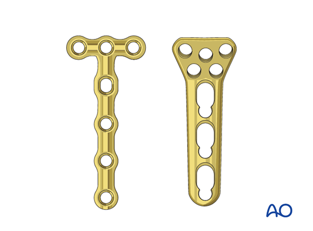
As the radial head is a small fragment, a 1.5 or 2.0 T-plate, or a locking proximal radius plate is used to allow purchase of two or three screws in the proximal fragment. Using locking screws may be safer as there is no need to have bicortical fixation.
The plate should be long enough to allow insertion of three screws in the distal fragment.
Pre-bend the plate according to the surface anatomy of the proximal radius. A slight convex bend at the fracture site may improve compression of the opposite cortex.
5. Creating compression
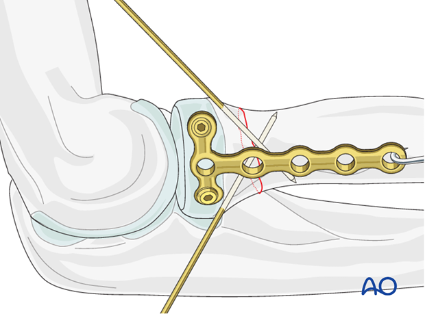
Attach a small bone hook in the distal hole of the plate and apply distal traction to the plate creating compression at the fracture site. At the time of application of compression, the temporary K-wires could be removed to enhance the compression effect. In this case use a small clamp to stabilize the fracture.
Insert two or three screws (1.5 mm or 2.0 mm) through the plate into the proximal fragment. These are parallel to the proximal surface and entirely within the bone.
6. Final assessment
Finally, assess the range of motion in pronation, supination, flexion and extension. Fixation should be stable and crepitus or restricted motion should be absent. Radiocapitellar and ulnohumeral joints should remain located through a full range of motion.
Check fractures and implants with image intensifier or x-ray.
If possible, close the annular ligament with non-resorbable sutures.
