Medial: Taylor and Scham
1. Introduction
The Taylor and Scham approach is a good choice for medial plate fixation of large, basilar fractures of the coronoid.
2. Medial approaches
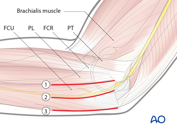
The interval that splits the flexor pronator mass and elevates the anterior part (pronator teres (PT), flexor carpi radialis (FCR), and palmaris longus (PL)) along with brachialis from the anterior elbow capsule gives good access to the anterior elbow capsule and the tip of the coronoid also known as the Hotchkiss “over the top” approach (1). The access to the medial facet of the coronoid is limited and there is also poor access to the base of the coronoid.
For access to the medial facet, use the interval where the ulnar nerve lies between the heads of the flexor carpi ulnaris (FCU split; 2).
For access to the base consider elevating the entire flexor-pronator mass from posterior to anterior (Taylor and Scham; 3).
In the following the Taylor and Scham approach is described.
Skin incision
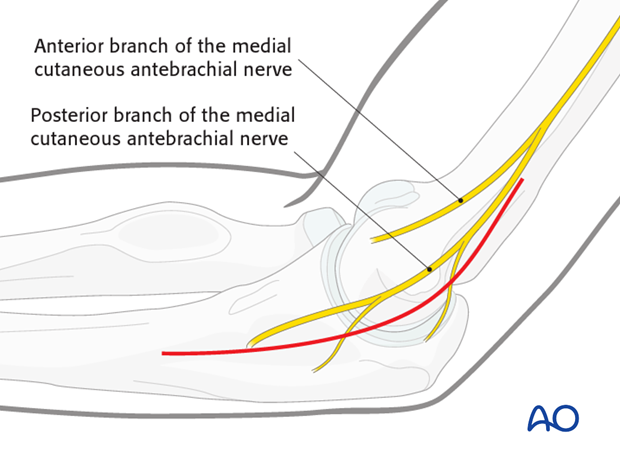
The skin incision can either be posterior with a medial skin flap or direct medial, taking care to protect the posterior branches of the medial antebrachial cutaneous nerve
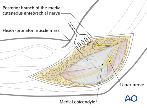
Ulnar nerve
The ulnar nerve should be identified and protected. Generally, it is unroofed for 6 centimeters proximal and distal to the epicondyle. Consider anterior subcutaneous transposition if you think this will keep the nerve safer.
Pearl: Always start with the exposure of the ulnar nerve proximally as it is easier and safer.
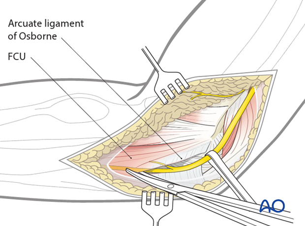
Follow the ulnar nerve distally as it goes under the fascia between the two heads of the flexor carpi ulnaris (FCU). Incise it with a pair of scissors and protective the first motor branch running to the humeral part of the FCU.
Pearl: Small, bleeding vessels can be best coagulated by using a bipolar coagulation pincette to protect the ulnar nerve.
Muscle elevation
Elevate the FCU and the entire flexor-pronator mass extraperiosteally from posterior to anterior starting at the crest of the ulnar shaft and the flat surface of the olecranon using a blunt elevator. You should expose the base of the coronoid fracture. If the ulnar nerve is at risk, transpose it anteriorly into the subcutaneous tissues.
Pearl: It is crucial to preserve the medial collateral ligament. Sometimes it gets difficult to distinguish the tendinous origin of FCU from the fibers of the medial collateral ligament. It is helpful to dissect from distal to proximal towards the sublime tubercle which is usually palpable. As long as the dissection is extra-periosteal and only muscle is elevated from the bone, the ligament should be safe.
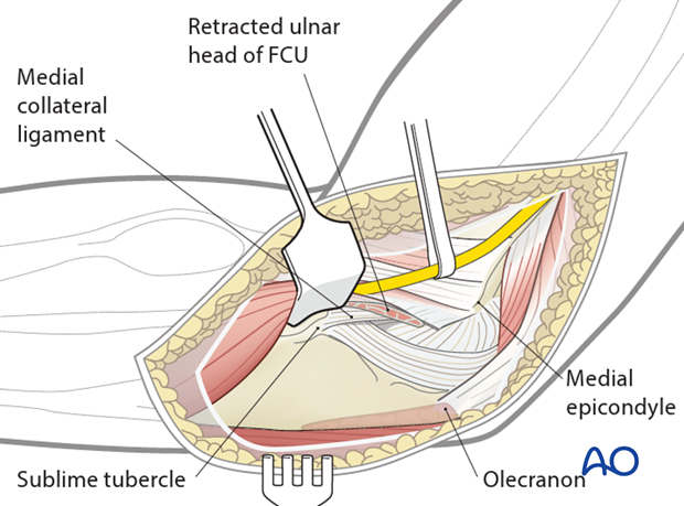
If access to the anterior capsule is needed, make a second more anterior interval such as the “over the top” exposure. Elevation of the origin of the flexor-pronator mass off the medial supracondylar ridge of the distal humerus, gives good exposure, but is destructive and should be avoided if possible.


