Lateral approach to the proximal forearm
1. Introduction
The lateral Kocher/Kaplan approach can be used to access the radial head and the tip of the coronoid.
2. Skin incision
Either a posterior skin incision with a lateral skin flap or a lateral skin incision can be used.
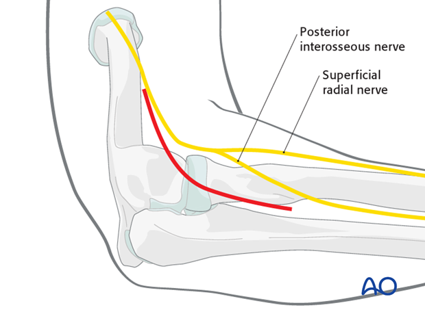
For a lateral skin incision, place the elbow at 90 degrees and try to pinch the lateral condyle (easier in thin patients). Make a straight skin incision directly over the middle of the lateral condyle. Start with a small incision (6-8 cm or so) and extend proximal or distal as needed.
Note: The posterior interosseous nerve, within the supinator muscle, crosses the posterior radius, from anteriorly, three finger-breadths distal to the radial head. It must be protected during this approach.
3. Superficial surgical dissection
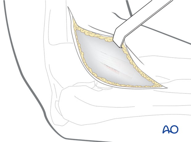
Incise the subcutaneous tissue in line with the incision and raise flaps to expose the fascia over the muscles.
4. Kaplan interval
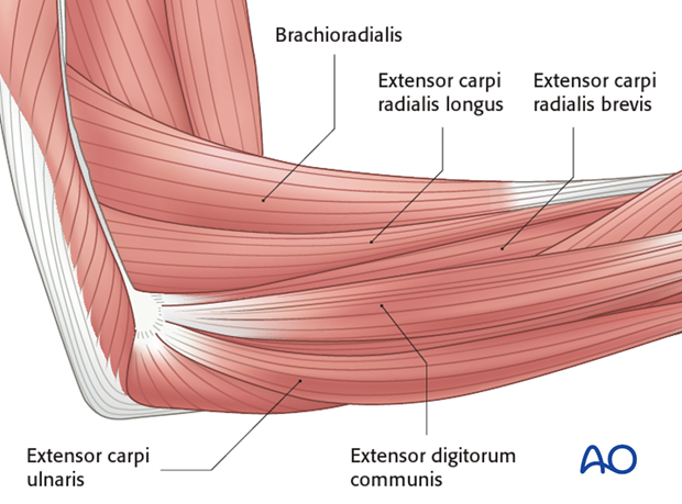
It can be difficult to determine exact muscle intervals. When we are operating on the radial head, there is usually associated injury to the lateral collateral ligament and common digit and wrist muscle origins. Along with this damage there is usually a rent in the fascia that can be opened and extended distally. This usually lies in the interval between the extensor carpi radialis brevis and extensor digitorum communis (Kaplan interval). The associated ligament and muscle injury will make the rest of the exposure very easy.
The Kaplan interval can also be identified by elevating the origin of the extensor carpi radialis brevis from the supracondylar ridge of the distal humerus, elevating the brachialis from the anterior humerus, then continuing distally until the joint is entered and the capitellum is visualized. Elevating these muscles is necessary to exposure the coronoid from the lateral side. Split the common wrist and digital extensor musculature at the point that divides the capitellum in half anterior/posterior.
5. Kocher interval
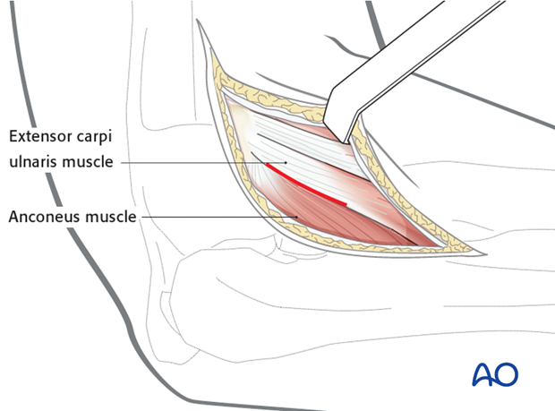
The interval between the anconeus and extensor carpi ulnaris (Kocher interval) is relatively more posterior and thus risks injuring the lateral collateral ligament complex.
6. Avoiding damage to radial nerve
- Fully pronating the forearm protects the posterior interosseous nerve by moving it away from the operative field.
- Beware of incising the capsule too far anteriorly as the radial nerve lies over the front of the anterolateral portion of the elbow capsule.
- Beware of dissection distal to the annular ligament or strenuous retraction, because the posterior interosseous nerve lying within the supinator muscle is at risk.
- No retractor should be placed around the radial neck.
7. Deep surgical dissection
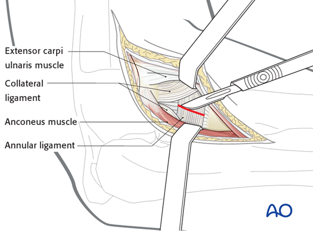
The following illustrations will show the deep dissection for the Kocher interval.
The annular ligament is divided in line with the muscle interval.
8. Osteotomy of lateral epicondyle
If the lateral collateral ligament is intact and better exposure of the radial head and neck are desired, one can consider osteotomy of the lateral humeral epicondyle. The osteotomy line in the illustration is marked in red.
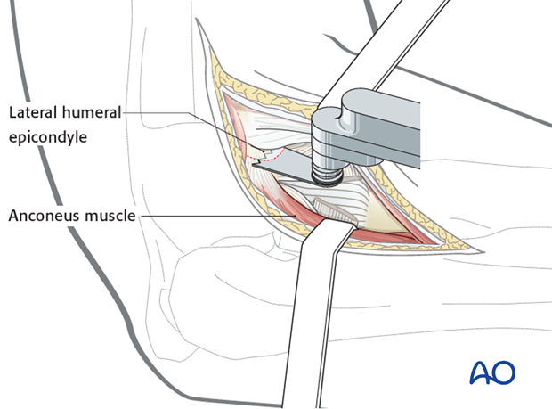
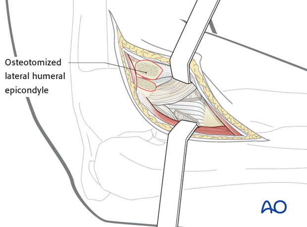
The soft tissues and osteotomized lateral epicondyle are reflected anteriorly to provide access to the proximal radius and ulna.
Screw fixation of the osteotomy can be difficult because the fragment is small and metaphyseal. A cerclage wire is an alternative, using the muscle origin as a more reliable point of fixation.
9. Closure
The wound is closed in layers.


