Multifragmentary pertrochanteric fractures with incompetent lateral wall
Definition
Multifragmentary pertrochanteric fractures with an incompetent lateral wall are classified by AO/OTA as 31A2.
The coronal fragment may be further fractured.
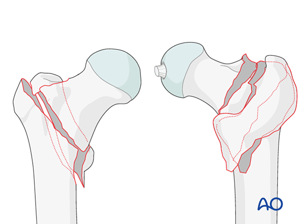
Lateral wall height of greater trochanter
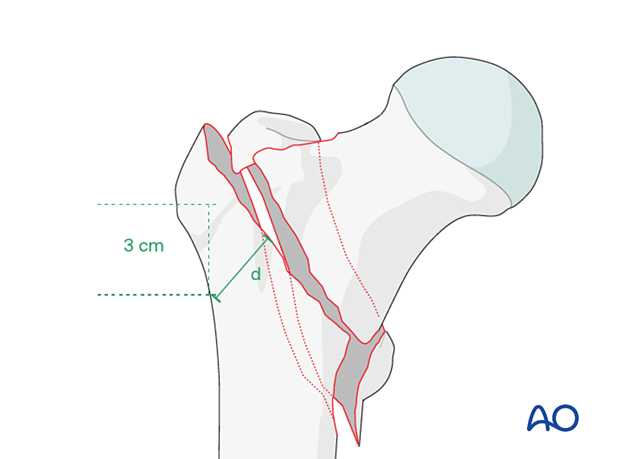
The coding system separates the pertrochanteric fractures into two groups (A1 and A2) defined by the lateral wall height (d) of the greater trochanter.
Lateral wall height or thickness is defined as the distance in millimeters (mm) from a reference point 3 cm below the innominate tubercle of the greater trochanter angled 135° upward to the fracture line on the AP x-ray. The thickness (d) must be less than 20.5 mm for the fracture to be considered an A2 fracture (Hsu et al 2013). It is recommended that the measurement for the lateral wall be taken using the traction view with the leg in neutral rotation. This can be difficult to obtain preoperatively but should be assessed fluoroscopically following operative closed reduction but before implant selection. Alternatively, preoperative 3-D CT provides detailed mapping of fracture location and planes.
Further characteristics
Fractures with an incompetent lateral wall (≤20.5 mm) are unstable after reduction. The greater trochanter is involved and fractured, and often displaced.
Multifragmentary pertrochanteric fractures may be unstable, particularly if the number of intermediate fragments is larger. Stability is also reduced if the greater trochanteric mass is involved in the (four-part) fracture or the fracture line extends well (>1 cm) below the lesser trochanter.
The exact fracture pattern is often difficult to determine on emergency x-rays. 3-D CT can provide this information.
In extracapsular fractures, there is minimal risk of osteonecrosis of the femoral head.
Additional coronal fragment
In addition to a primary trochanteric fracture, often a secondary coronal fracture on the posterior aspect of the greater trochanter can be observed. These coronal fragments may start at the summit of the greater trochanter and exit through some point along the trochanteric crest or even at the posteromedial cortex.
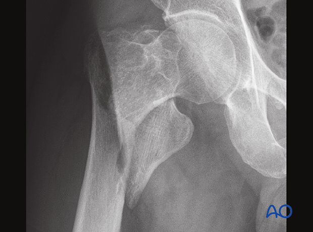
Case of a 31A2.2 fracture (with one intermediate fragment)
This case shows a three-part fracture with an incompetent lateral wall and a big fragment, including the lesser trochanter.
In the CT scan of the same case, the distal fracture extension of the coronal fragment is clearly visible.
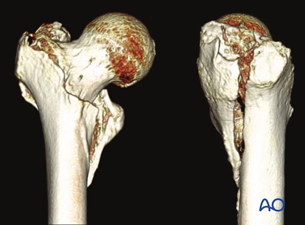
It includes the lesser trochanter.
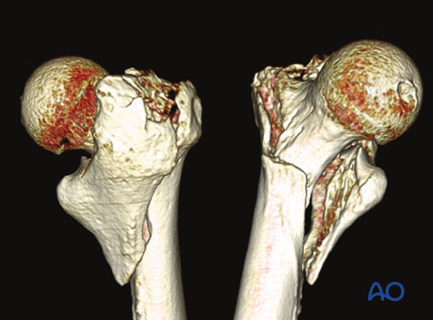
Case of a 31A2.3 fracture (with two or more intermediate fragments)
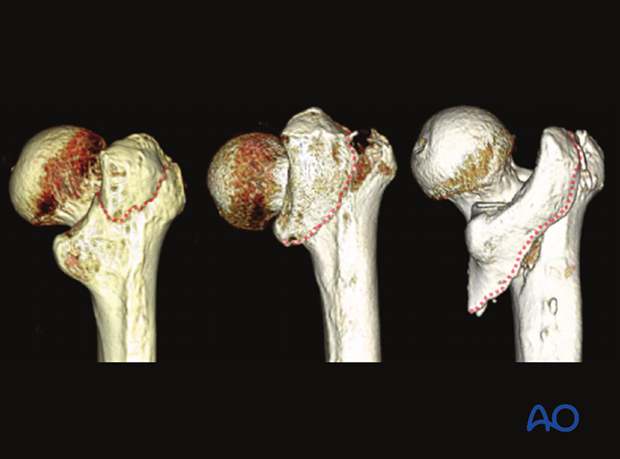
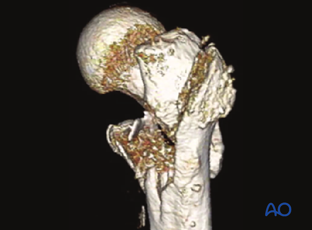
This case shows the primary fracture line along the trochanteric crest with an incompetent lateral wall and a large posteromedial fragment.
It is difficult to obtain the detailed fracture morphology based on this x-ray only.
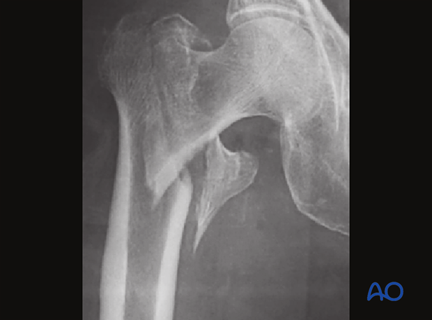
In the anterior view of the 3-D CT, the primary fracture line can be recognized.
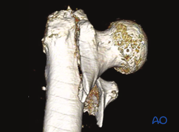
On the posterior view, the coronal fragment is visible, showing detachment of the posteromedial fragment including the lesser trochanter.
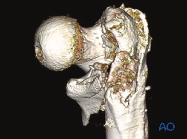
Lateral view of the same case
Occult hip fractures
An undisplaced fracture may also be referred to as occult fracture as it is often not visible and may not be diagnosed correctly.
If clinical assessment indicates a neck fracture, but the x-ray does not show clear signs of it, CT or MRI imaging is recommended.


