Hook plate fixation
1. General considerations
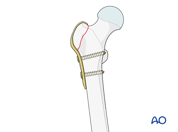
If implants are available, hook plate fixation is the preferred fixation technique. It provides good stability in osteoporotic bone.
A short plate with two holes for screw fixation in the subtrochanteric area is sufficient.
2. Patient preparation and approach
Patient positioning
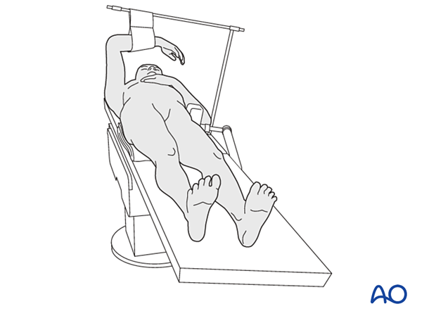
Place the patient supine on a radiolucent table with a bump under the buttock.
Approach
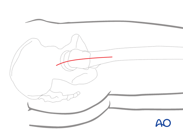
For this procedure, a direct lateral approach centered over the tip of the greater trochanter is used.
3. Reduction
Clear any hematoma.
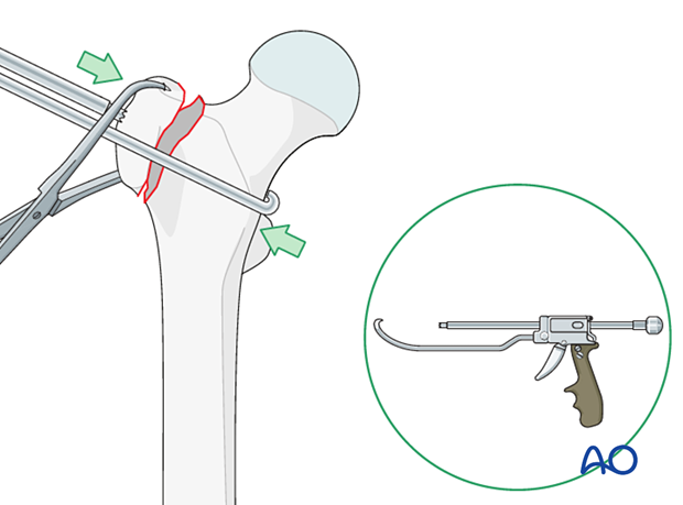
Directly reduce the fragment and stabilize it with pointed bone reduction forceps, one anteriorly and one posteriorly, leaving space for plate application. Abducting the extremity facilitates reduction.
The collinear reduction forceps may be helpful to reduce and stabilize the fracture. It is applied from the direct lateral incision with proximal extension. It needs to be replaced by two reduction forceps to give space for plate application.
4. Plate application
Apply the plate on the lateral side of the femur to the proximal segment through the abductor tendon.
Perform separate stab incisions through the gluteus medius and place the two hooks into the tip of the greater trochanter.
Confirm with image intensification that the hooks have a solid purchase in the trochanter.
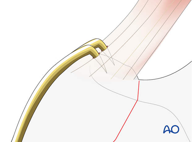
Insert cortical screws bicortically to fix the plate to the proximal shaft.

5. Final assessment
Confirm complete reduction, stability, and range of motion.
Obtain AP and lateral x-rays to confirm correct implant position.
6. Aftercare
Postoperative mobilization
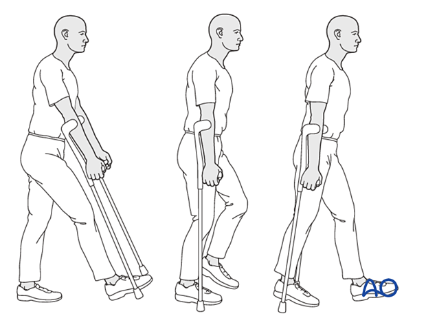
Weight-of-the-leg weight bearing with walking aids will decrease the pull of the abductors on the fragment and is recommended for 4–6 weeks.
Pain control
To facilitate rehabilitation and prevent delirium, it is important to control the postoperative pain properly, eg, with a specific nerve block.
VTE prophylaxis
Patients with lower extremity fractures requiring treatment require deep vein prophylaxis.
The type and duration depend on VTE risk stratification.
Follow-up
Follow-up assessment for wound healing, neurologic status, function, and patient education should occur within 10–14 days.
At 3–6 weeks, check the position of the fracture with appropriate x-rays.
Recheck 6 weeks later for progressive fracture union.


