Intertrochanteric fractures
Definition
Intertrochanteric fractures are classified by AO/OTA as 31A3. They are often called reverse oblique fractures.
These are true intertrochanteric fractures. The fracture line passes between the two trochanters, above the lesser trochanter medially and below the crest of the vastus lateralis laterally. Both femoral cortices are involved.
This fracture type is subdivided:
- 31A3.1 – Simple oblique fracture
- 31A3.2 – Simple transverse fracture
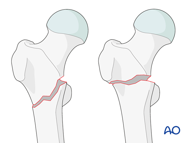
- 31A3.3 – Wedge or multifragmentary fracture
The most common fracture type is 31A3.3 multifragmentary.
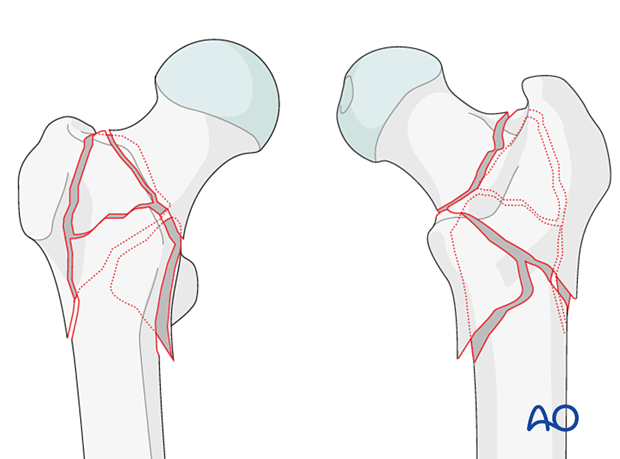
Most of the time, there is coronal fragmentation. This can involve the greater trochanter down to the posteromedial cortex and may even be further fragmented.
Further characteristics
Additional coronal fragment
In addition to a primary trochanteric fracture, often a secondary coronal fracture on the posterior aspect of the greater trochanter can be observed. These coronal fragments may start at the summit of the greater trochanter and exit through some point along the trochanteric crest or even at the posteromedial cortex.
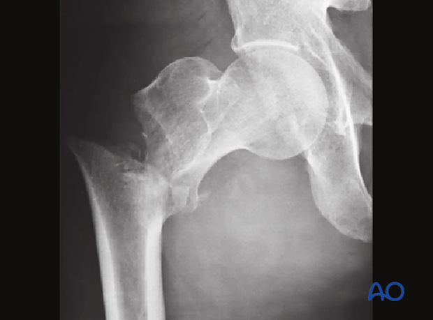
Simple oblique fracture (31A3.1)
AP x-ray and 3-D CT views
Reverse oblique fracture
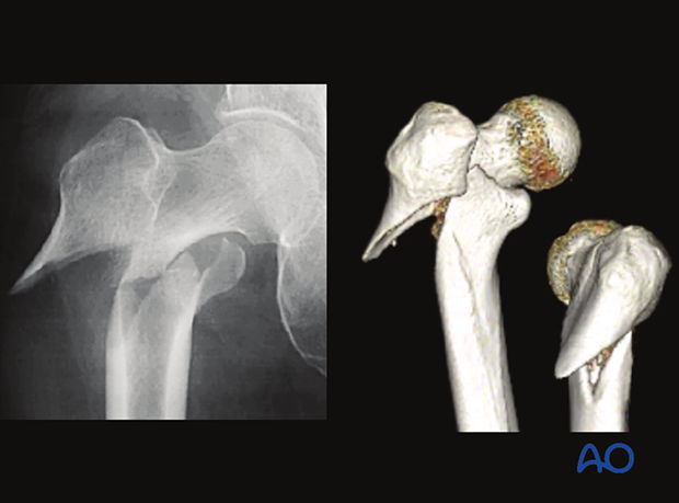
The so-called reverse oblique fractures often have a typical displacement because of the pull of the abductors, which abduct and flex the proximal fragment. Be careful in determining the extent of the fracture, as undisplaced fissures down into the femoral shaft and up into the trochanteric block are common.
Low lateral escape fracture
This simple intertrochanteric fracture is referred to as low lateral escape fracture. The fracture line starts near the vastus ridge of the greater trochanter. This pattern is referred to as the low lateral escape fracture.
Typically, the fracture plane of trochanteric fractures is nearly perpendicular to the direction of the sliding. In the low lateral escape fracture, nailing can not provide sliding because both the nail head and blade/lag screw are in the proximal fragment. Without anatomical reduction, nonunion may occur.
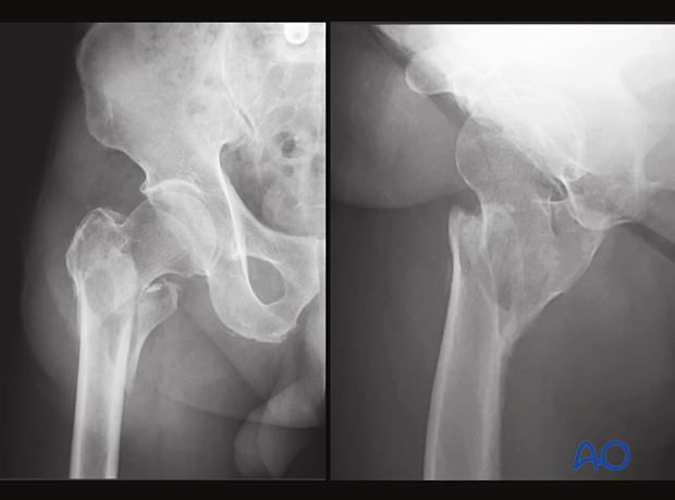
Multifragmentary intertrochanteric fracture (31A3.3)
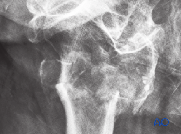
Plain AP and lateral views of a multifragmentary fracture
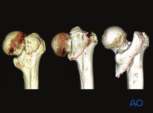
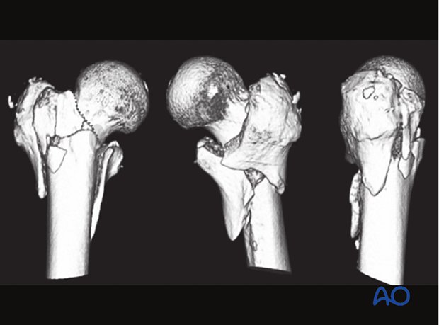
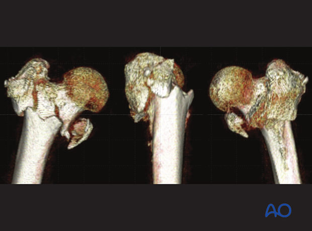
Coronal fragments are much better recognized on 3-D CT than on plain x-rays.
This 3D-CT shows two coronal fragments, ie, the greater trochanter and posteromedial cortical fragment.
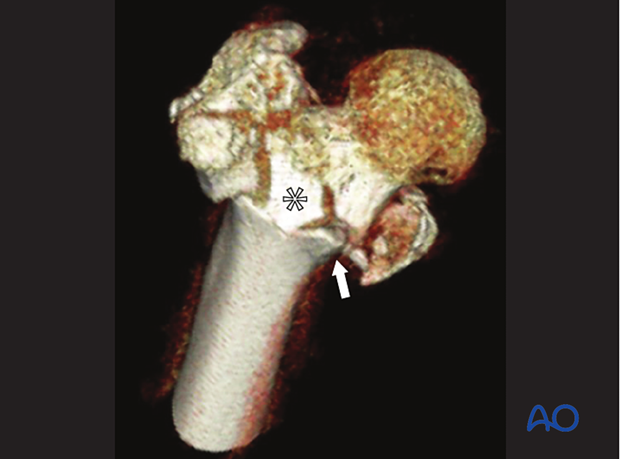
Case with an additional anterior fragment
AP x-ray of a multifragmentary intertrochanteric fracture
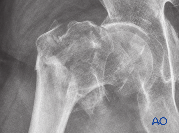
Lateral view
In the 3-D CT, the fragmentation pattern becomes more readable and helps to understand the fracture personality, decision making, and preoperative planning.
In this case, the anterior cortex contact is very limited (arrow) due to an additional anterior fragment (asterisk).
Occult hip fractures
An undisplaced fracture may also be referred to as occult fracture as it is often not visible and may not be diagnosed correctly.
If clinical assessment indicates a neck fracture, but the x-ray does not show clear signs of it, CT or MRI imaging is recommended.


