Supine patient position for arthroplasty
1. Indications
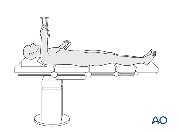
Supine patient positioning is used for hemi and total hip arthroplasty. The use of a radiolucent table allows for intraoperative assessment of acetabular cup position and the length of the prosthesis.

2. Preoperative preparation
Operating room personnel (ORP) need to know and confirm:
- Site and side of the fracture
- Type of operation planned
- Ensure that the surgeon has marked the operative site
- Condition of the soft tissues (fracture: open or closed)
- Implant to be used
- Patient positioning
- Details of the patient (including a signed consent form and appropriate antibiotic and thromboprophylaxis)
- Comorbidities, including allergies
3. Perioperative care for elderly hip-fracture patients
Routine perioperative care includes:
- Brief intravenous antibiotics
- VTE prophylaxis (before, during, and after surgery)
- Nutritional supplementation
- Pain management without oversedation
- Prevention of pressure sores
- Early mobilization
- Early discharge planning
- Osteoporosis evaluation and management
For more details, see the additional material on perioperative care for elderly hip fracture patients.
4. Anesthesia
This procedure is performed with the patient under general or regional anesthesia.
5. Patient positioning
Place the patient supine and as close as possible to the edge of the table.

6. C-arm positioning
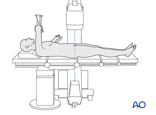
Position the C-arm perpendicular to the table. AP views can be obtained easily.

7. Intraoperative options for VTE prophylaxis
During the operation, some centers provide VTE prophylaxis with sequential mechanical compression on the contralateral leg.
8. Skin disinfection and draping
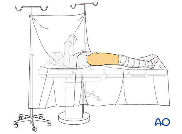
Disinfect the exposed area from above the iliac crest to the mid-tibia with the appropriate antiseptic solution.
Free drape the affected limb(s) with a single-use U-drape. A stockinette covers the lower leg and is fixed with tape. The leg is draped to be freely moved.
Drape the image intensifier.

9. Operating room set-up
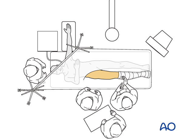
The surgeon, assistant, and ORP stand on the side of the injury.
Place the image intensifier on the opposite side of the injury or surgeon.
Place the image intensifier display screen in full view of the surgical team and the radiographer.


