Transcervical shear fractures of the femoral neck
Definition
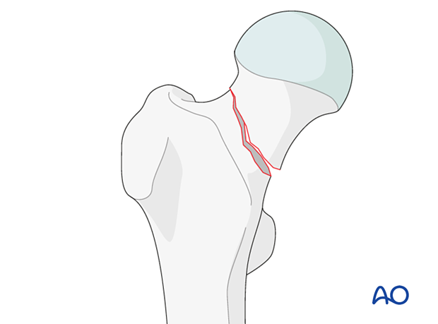
Transcervical shear fractures of the femoral neck are classified by AO/OTA as 31B2.3.
The inclination of the fracture line on the AP view is more than 50° from the horizontal (Pauwels type III).

Further characteristics
These are extraarticular but intracapsular fractures.
The blood supply to the femoral head is threatened because the displacement may disrupt the blood vessels leading to the femoral head.
This fracture type is very unstable due to the shearing forces.
The blood supply of the femoral head
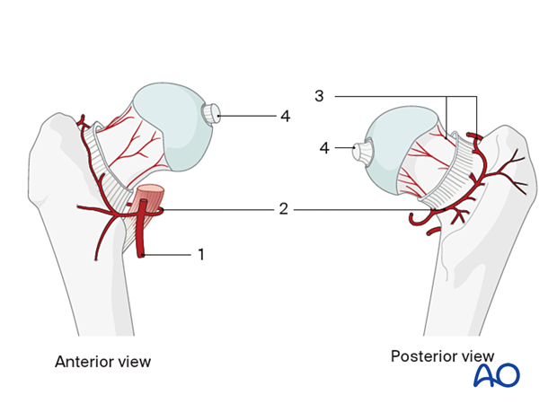
The vascular anatomy varies, but in 60% of patients, the medial and lateral femoral circumflex arteries originate from the profunda femoris artery (1).
Most of the blood supply of the femoral head comes from the medial femoral circumflex artery (2), which gives rise to three or four branches, the retinacular vessels (3). These run posteriorly and superiorly along the femoral neck in a synovial reflection until they reach the cartilaginous border of the head. The obturator artery gives rise to the vessels within the ligamentum teres (4). The medial and lateral circumflex arteries may anastomose, but the principal blood supply of the head originates from the medial circumflex artery and its branches, especially the ascending ones.

Imaging
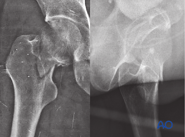
In the AP view, there is a disruption of the medial and lateral cortical line and a vertical fracture line.
In the lateral view, there may be a step-off in the anterior cortical line.


