Impacted or nondisplaced subcapital femoral neck fractures
Definition
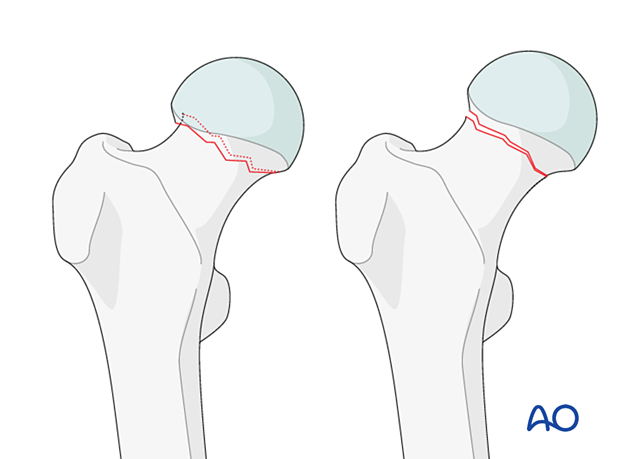
Subcapital femoral neck fractures with valgus impaction or with little or no displacement are classified by AO/OTA as 31B1.1 and 31B1.2, respectively.
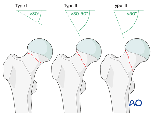
The Pauwels classification can best be determined intraoperatively, once traction is applied and the fracture is reduced. On an AP fluoroscopic view, assess the inclination of the fracture line. Pauwels type I is a fracture with <30º from the horizontal. Type II is 30º–50º from the horizontal. Type III is >50º from the horizontal.
Further characteristics
In these fractures, the contact between the head and the neck is always maintained.
The undisplaced fractures are unstable before fixation since there is no impaction.
Neck fractures are extraarticular but intracapsular; the articular surface is not damaged, but the blood supply to the femoral head may be compromised.
Impacted fractures are usually caused by a fall of an elderly patient to the side. In younger patients, this fracture is very unlikely.
Imaging
X-rays
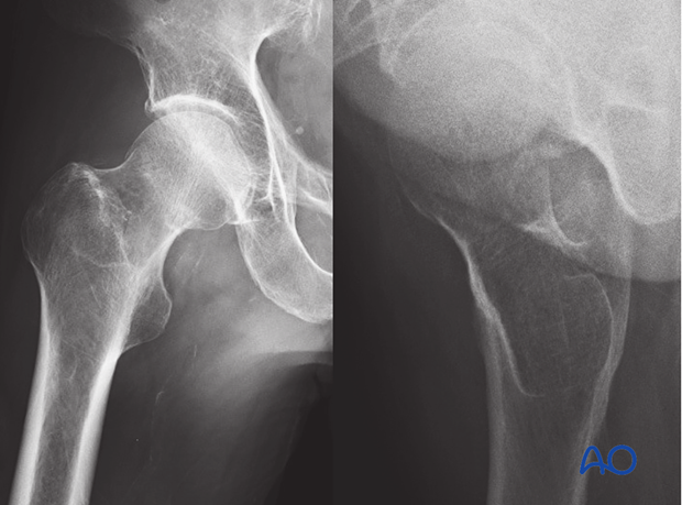
On the AP x-ray of an impacted fracture, the lateral cortex of the head-neck junction is overlapped.
On the lateral view, there will be normal anteversion.
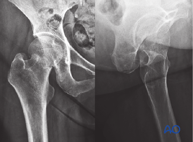
In the AP x-rays of an undisplaced fracture, the fracture line is visible, but both the medial and lateral cortical lines of the neck show no gap. The “lazy” S-sign is intact.
In the lateral view, there should be normal anteversion.
Occult hip fractures
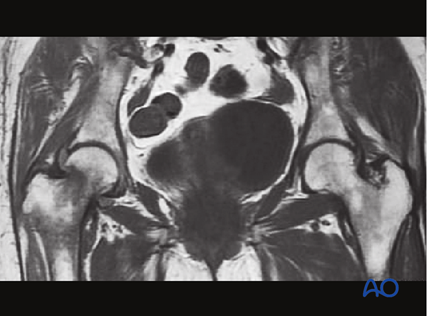
An undisplaced fracture may also be referred to as occult fracture as it is not well visible in an x-ray and may not be diagnosed correctly.
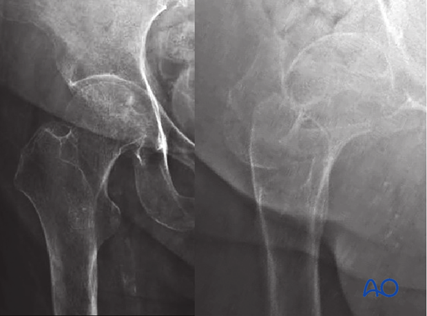
If clinical assessment indicates a neck fracture, but the x-ray does not show clear signs of it, CT or MRI imaging is recommended (Davidson et al 2020).
