Femoral neck and head fractures with hip dislocation
Definition
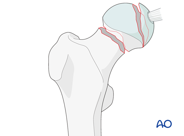
Combined fractures of the femoral head and neck are uncommon and associated with dislocations of the hip.
They are caused by very high-energy traumas.

Further characteristics
Because these fractures involve the femoral head and neck, the blood supply to the fracture fragments is almost always disrupted.
The blood supply of the femoral head
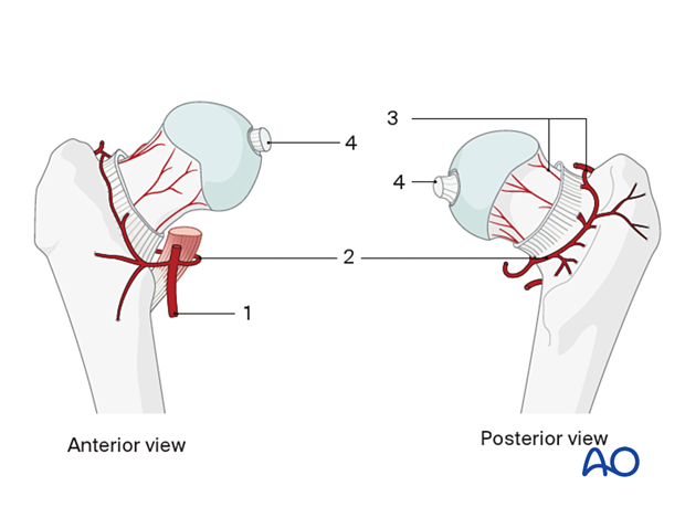
The vascular anatomy varies, but in 60% of patients, the medial and lateral femoral circumflex arteries originate from the profunda femoris artery (1).
Most of the blood supply of the femoral head comes from the medial femoral circumflex artery (2), which gives rise to three or four branches, the retinacular vessels (3). These run posteriorly and superiorly along the femoral neck in a synovial reflection until they reach the cartilaginous border of the head. The obturator artery gives rise to the vessels within the ligamentum teres (4). The medial and lateral circumflex arteries may anastomose, but the principal blood supply of the head originates from the medial circumflex artery and its branches, especially the ascending ones.

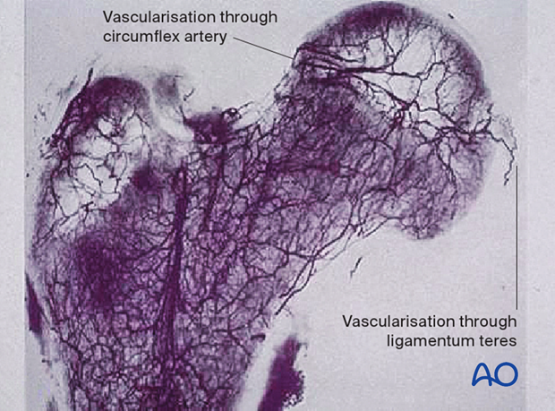
Vascularization through ligamentum teres
The ligamentum teres arises from the transverse acetabular ligament and the posterior inferior portion of the acetabular fossa and attaches to the femoral head at the fovea capitis. Lesions of the ligamentum teres may be caused by dislocation or subluxation of the hip as well as acetabular fractures.
However, the blood supply through the ligamentum teres is minor (10–15% of the femoral head) and mostly to the non-weight-bearing area.

Imaging
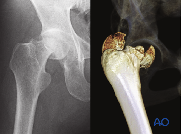
Good quality imaging (x-ray and CT) is essential for the diagnosis and planning of the treatment.


