Depression fractures of the femoral head
Definition
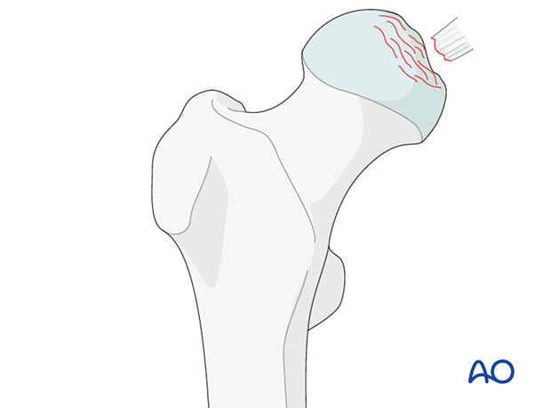
Depression fractures of the femoral head are classified by AO/OTA as 31C2.
This fracture type is subdivided:
- 31C2.1 – Chondral lesion
- 31C2.2 – Depression impaction fracture
- 31C2.3 – Split depression fracture
They are usually associated with acetabular fractures and should be suspected in every patient with an acetabular fracture.
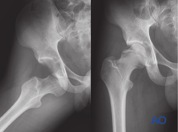
Depression fractures are also associated with anterior hip dislocation.
Combined with a depression fracture of the femoral head, there might also be an avulsed fragment.

Further characteristics
These fractures result from high-energy trauma and are rare.
The injury to the femoral head is often not evident until visualization of the head at surgery. Because of the technical surgical difficulties in dealing with these fractures, they demand a surgeon with special expertise in pelvic and acetabular trauma.
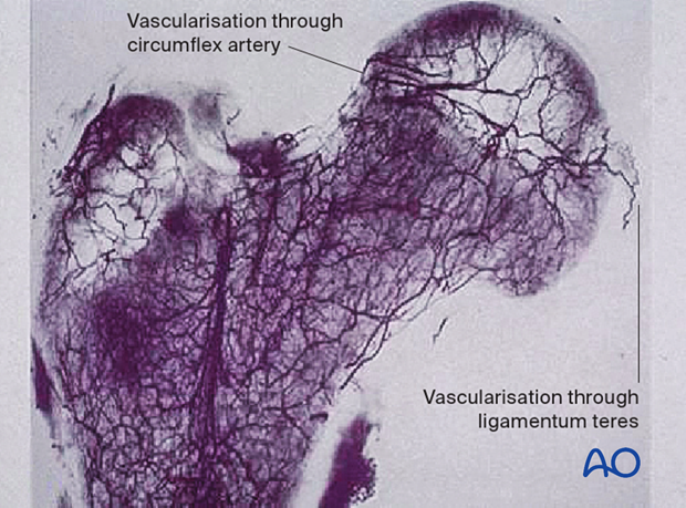
Vascularization through ligamentum teres
The ligamentum teres arises from the transverse acetabular ligament and the posterior inferior portion of the acetabular fossa and attaches to the femoral head at the fovea capitis. Lesions of the ligamentum teres may be caused by dislocation or subluxation of the hip as well as acetabular fractures.
However, the blood supply through the ligamentum teres is minor (10–15% of the femoral head) and mostly to the non-weight-bearing area.

Imaging
A CT scan should be obtained to determine the morphology of the fractures.
An MRI may add additional information but is not sufficiently sensitive to diagnose articular cartilage lesions.


