Prone
1. Indications
The prone position is used for access to the sacrum and posterior SI joint regions.
2. Positioning
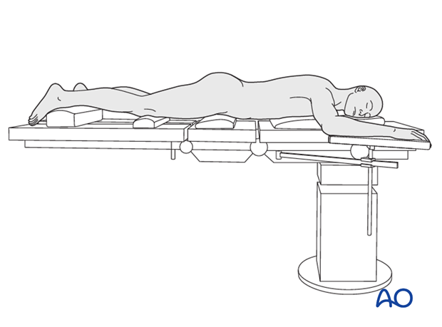
Place the patient prone on the radiolucent operating table.
Position bolsters to support the chest wall and pelvis; the bolsters should allow the chest wall and abdomen to expand without touching the table.
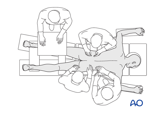
Take great care during preparation and draping to exclude the contaminated anal region from the operative field.
The knee on the injured side should be flexed to relax the sciatic nerve. All pressure points should be padded.

3. Radiolucent table
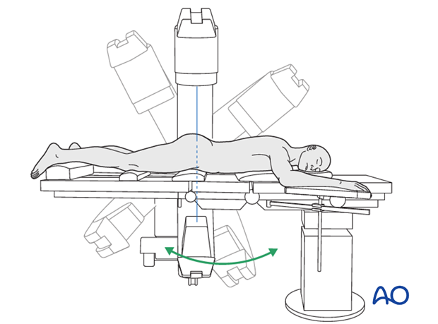
Assure that adequate images can be obtained (anterior-posterior, inlet and outlet) before draping the patient.
Prior to draping, ensure that the C-arm is able to obtain the necessary images without being obstructed by the table.

4. Prone with fracture table
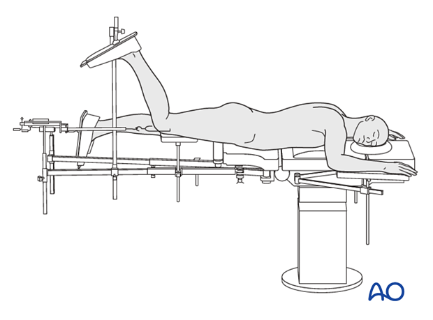
Alternatively, the patient can be positioned prone on an appropriate fracture table. IT is important that support post of the fracture table is close to the head end. This makes it possible to obtain adequate inlet and outlet views.
Such tables permit skeletal traction, which is applied to the distal femur. The knee must be kept flexed, with an appropriate foot support, to relax the sciatic nerve.
Prior to draping, ensure that the C-arm is able to obtain the necessary images without being obstructed by the table.

The patient’s arms should be positioned and padded to minimize tension on the brachial plexus, and to allow access for the surgical team.


