Pubic ramus plate
1. Introduction
Fractures of the pubic rami are nearly always associated with further pelvic ring injuries. The treatment of these fractures is guided by the displacement and other associated pelvic ring injuries.
In many cases, a strong periosteum, the inguinal ligament and the pectineal ligament will provide adequate stability and no additional treatment is required. With more complex, unstable pelvic ring injuries, ramus fractures may require fixation to restore stability and promote healing.
The decision for operative vs. nonoperative is best made by evaluating the entire pelvic ring and its stability instead of focusing only on the ramus fracture.
For more complex injuries, posterior reduction and fixation must precede fixation of the ramus fractures. In these cases, the anterior fixation supplements and stabilizes the posterior fixation.
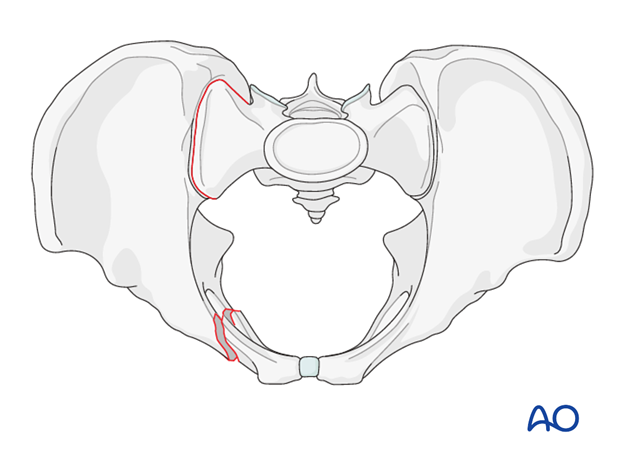
Options for fixation
Stabilization of ramus fractures can be performed by a variety of techniques:
- Internal fixation with an intramedullary screw
- Internal fixation with a plate
- Indirect stabilization with external fixation
For plate fixation, open anatomic reduction is desired. Typically, 3.5 mm reconstruction plates are utilized. Alternatively, any appropriate pelvic plate can also be used.
In osteoporotic bone and in comminuted fractures, angular stable plates are preferred.
The decision to use this technique is based on the pelvic injury pattern, nature of the ramus fractures, and the surgeon's comfort with the anatomy and imaging.
2. Open reduction
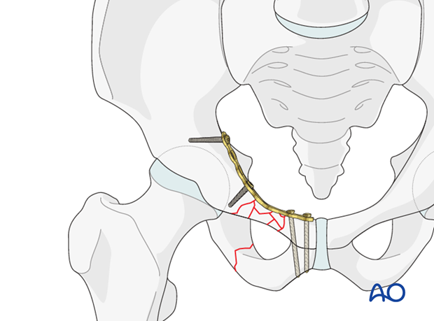
After adequate exposure of the fracture site is gained through an anterior approach, bone holding forceps are used to reduce the fracture by direct manipulation.
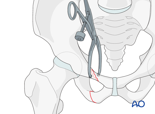
Reduction is verified by direct vision as well as with image intensifier using inlet and outlet views.
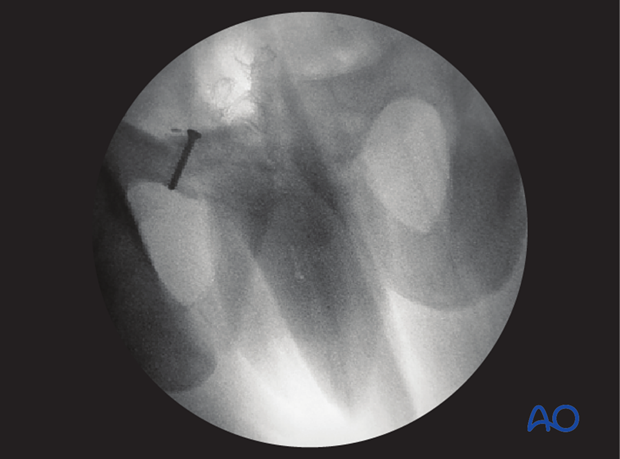
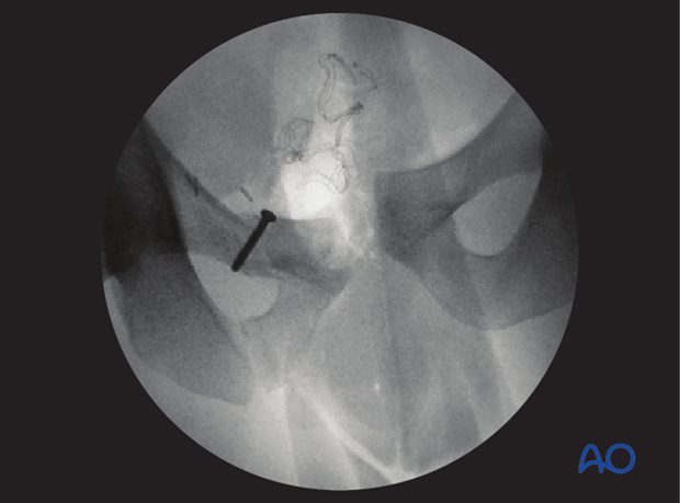
If anatomic reduction is not possible in comminuted fractures, a bridging technique with an appropriately contoured plate is used.
Note
Be alert for rotational malalignment of the fracture. This can be corrected by derotation by bone holding forceps.
3. Fixation
Fixation of oblique fractures
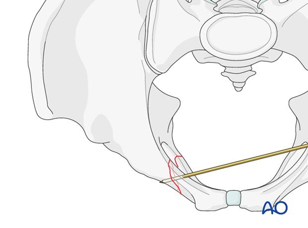
When anatomic reduction is achieved, K-wires are inserted perpendicular to the fracture plane to fix the fracture temporarily.
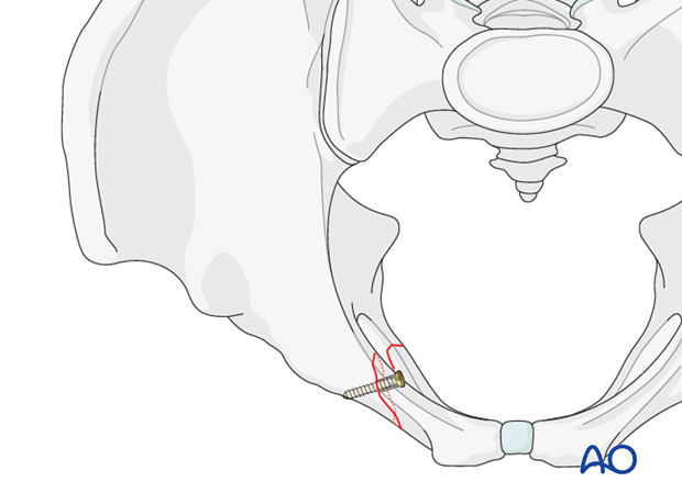
Insert the lag screw as perpendicular as possible to the fracture line.
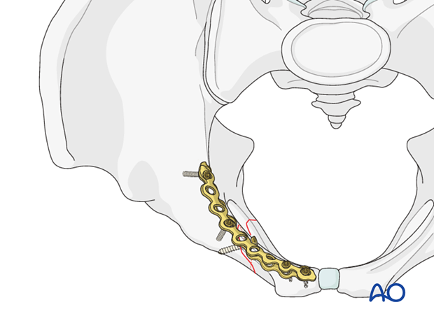
Place an appropriately contoured neutralization plate on the superomedial surface of the superior pubic ramus and fix it.
Fixation of transverse fractures
If the reduced fracture is stable, it will provide a template for appropriate contouring of the plate.
Remember to achieve compression of the transverse fracture by slightly underbending the plate which is being applied to a concave surface.
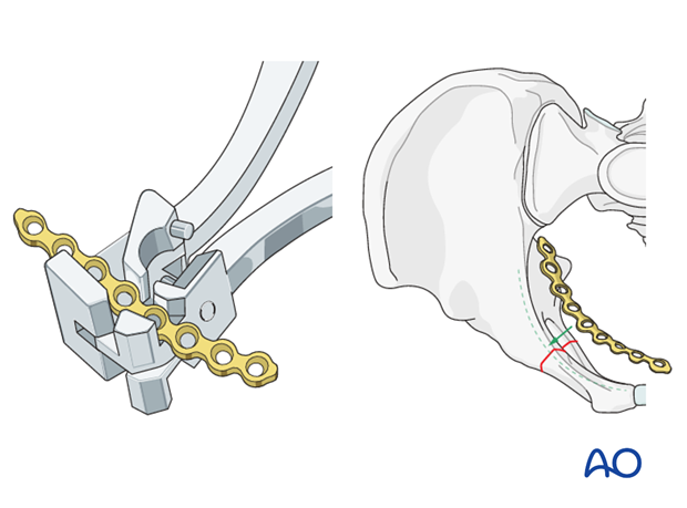
The plate is placed superomedial on the superior pubic ramus and inserted in compression mode.
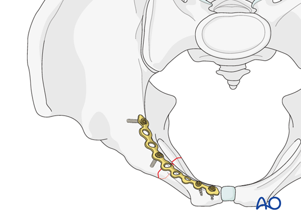
Fixation of comminuted fractures
In comminuted fractures it is important to evaluate the overall geometry of the pelvis.
When acceptable reduction of the pubic rami is achieved, an appropriately contoured plate is inserted as a bridging plate, superomedially on the superior pubic ramus.

Comment to medial and lateral fractures
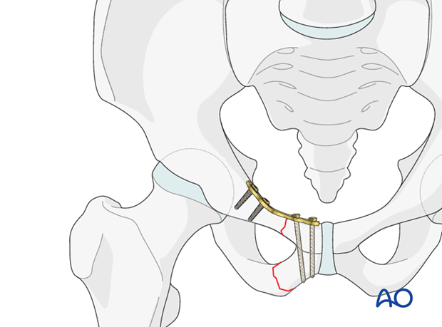
In medial fractures, it is possible to fix the plate to the ramus alone, both medially and laterally. At least two screws should be inserted on each side of the fracture line.
Extension of the plate across the symphysis to gain additional screws is possible if fixation is poor.
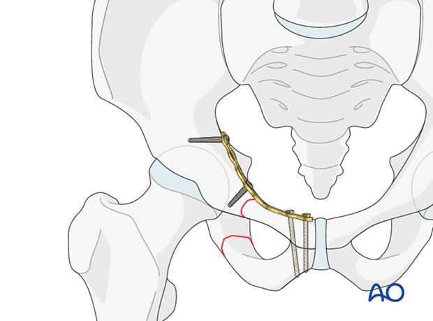
In lateral fracture patterns of the superior ramus, the lateral end of the plate should be fixed posterior to the acetabulum, as illustrated.
Verification of screw position
The position of each screw should be verified by C-arm to prevent screw penetration into the symphysis or the acetabulum.
Lateral screw positions are verified in the obturator and the iliac wing view.
The medial screws are verified using inlet and outlet views.
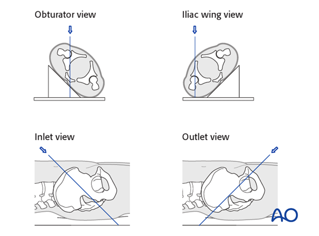
X-rays
After completion of internal fixation, confirm the final reduction and hardware position intraoperatively by AP, inlet and outlet radiographic imaging.


