Posterior approach to the sacrum
1. Indications
A posterior approach to the sacrum is indicated for exposure of its posterior surface and the posterior aspects of both SI joints, and adjacent posterior ilium.
It is typically used for ORIF of sacral fractures, especially with sacral nerve root decompression.
2. Skin incision
The skin incision varies according to the location of the fracture.
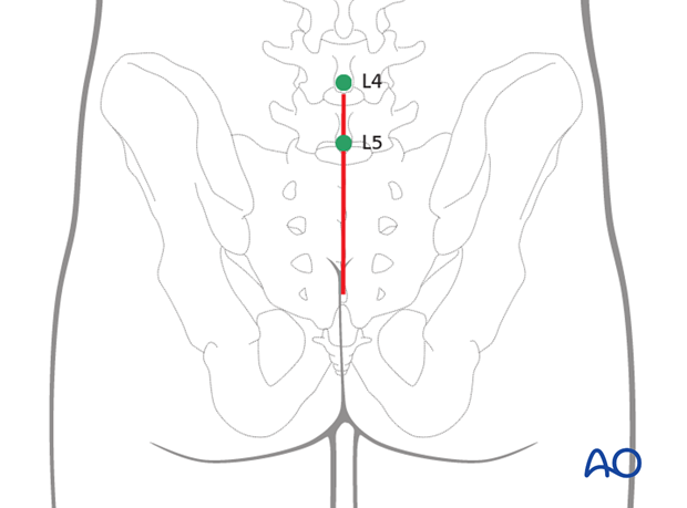
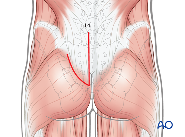
Use a midline incision to expose both sides of the sacrum.
The midline incision starts at the L4 or L5 spinous process and extends distally ending proximal to the anus.
Note: Posterior approaches through severely contused or crushed tissue or sites of subcutaneous hematoma (Morel-LaVallée lesions) may cause significant wound healing problems.
3. Alternative incision
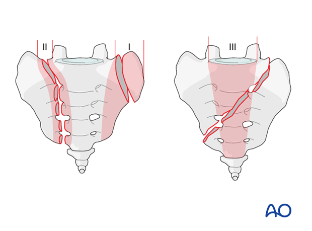
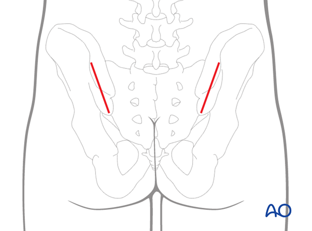
Incisions for transiliac fixation can be performed with oblique incisions starting at the posterior inferior iliac spine and a midline incision to allow submuscular implant insertion.
4. Transection of the muscle aponeurosis
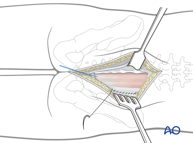
Incise the lumbosacral fascia parallel to the spinous processes L4 and L5 on the injured side.
A distally based V-shaped incision is made in the lumbosacral fascia.
Aim for the iliac crest and end the incision when this landmark is reached.
5. Dissection
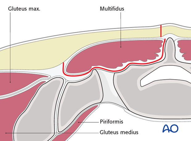
Subperiosteal elevation of the Iumbosacral musculature exposes the posterior aspect of the sacrum
This can be performed in one of two ways.
The first (as shown) involves releasing the multifidus distally and elevating it as a flap.
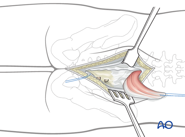
A second method involves incising the multifidus medially and laterally, elevating this muscle from the sacrum as a bi-pedicle flap.
If a more extensile exposure of the lateral sacral region („ala“) is necessary, elevate the complete muscle mass by dissecting its lateral attachment to the posterior iliac crest.
6. Wound closure
Initiate the closure of the wound by a gentle repositioning of the erector spinae muscles and reattachment of the superior origin of the gluteus maximus.
Close the subcutaneous tissues and skin in layers.
