Approach to the pubic symphysis
1. Exposure
The surgical approach to the anterior part of the pelvic ring is useful for:
- Pubic symphysis disruption/diastasis
- Fractures of the anterior pelvic ring including superior pubic rami
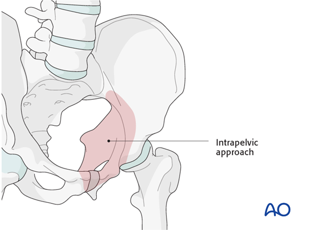
This approach may be carried laterally to expose the quadrilateral lamina (modified Stoppa approach).
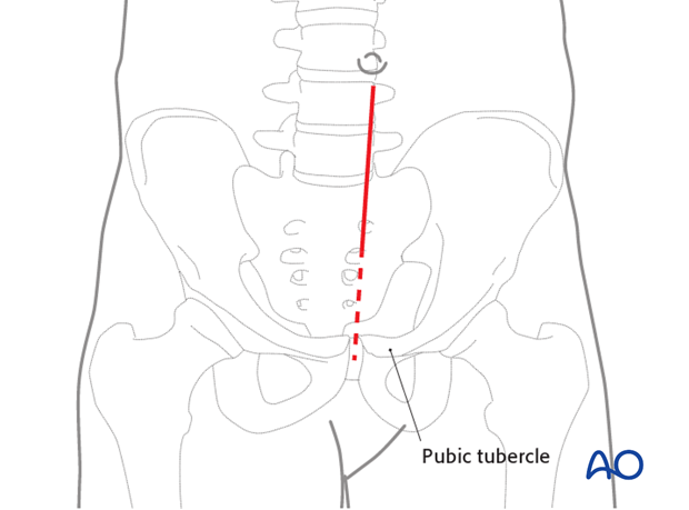
The illustrations show the portion of the anterior pelvic ring that can be exposed utilizing the standard approach.
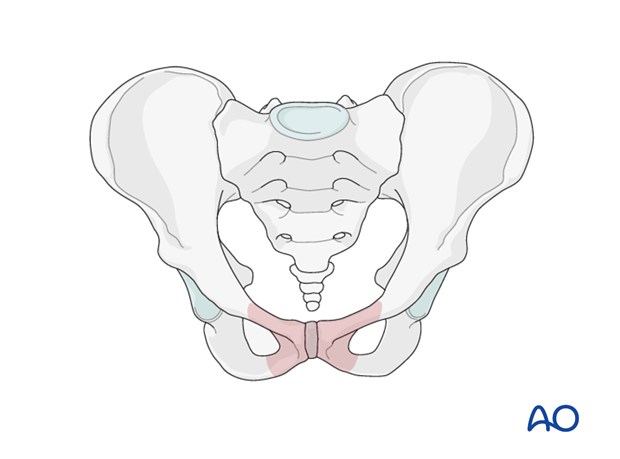
The approach can be extended laterally (modified Stoppa approach) to expose the anterior ring extending to the sacral iliac joint.
2. Horizontal incision (“Pfannenstiel”)
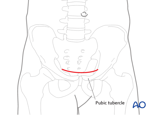
Perform a horizontal incision about 2 fingerbreadths proximal to the pubic tubercle.
The length of this incision is typically 5-10 cm. It can be extended further laterally on one or both sides depending on the needed exposure.
3. Vertical incision
If the patient has undergone a midline laparotomy, the inferior aspect of the laparotomy may be extended to provide exposure.
4. Dissection of subcutaneous tissue
Dissect the subcutaneous tissue and identify the anterior rectus fascia.
If the incision is extended laterally, identify and protect the spermatic cord or round ligament on both lateral sides of the wound.
Locate the linea alba and incise it longitudinally.
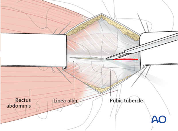
Both bellies of the rectus abdominis muscle are gently retracted laterally.
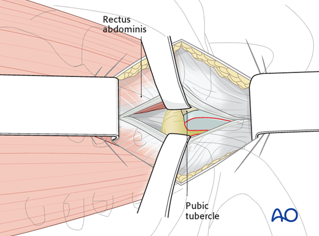
5. Exposure of the retrosymphyseal region
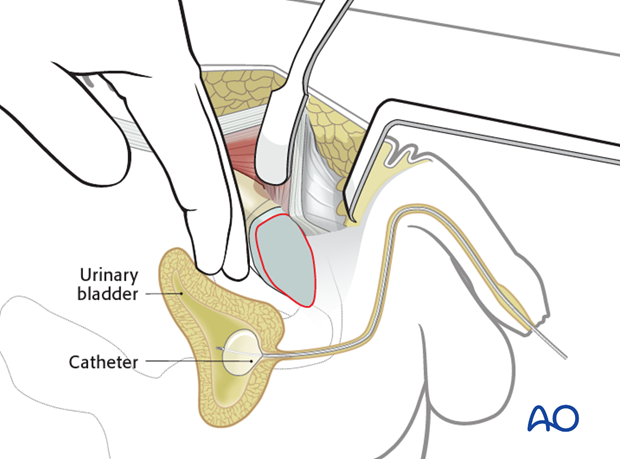
Use your hand to bluntly dissect the retro symphyseal region. Use care to avoid injury to the urinary bladder or to the prostatic venous plexus.
6. Retract the rectus abdominis
Usually in a symphyseal disruption, one side of the rectus abdominis is typically disrupted by the injury. This disruption usually results in the muscle insertion being avulsed from the pubis. In most cases, the rectus abdominis muscle remains in continuity.
Carefully elevate the remains of this disrupted rectus abdominis to allow exposure of the symphysis while maintaining as much distal fascial continuity as possible to avoid later hernia formation. Perform this dissection subperiosteally, extending laterally past the pubic tubercle.
Next, a pointed Hohmann retractor is placed obliquely over the pubic tubercle to retract the muscle and the distal aspect of the rectus abdominis.
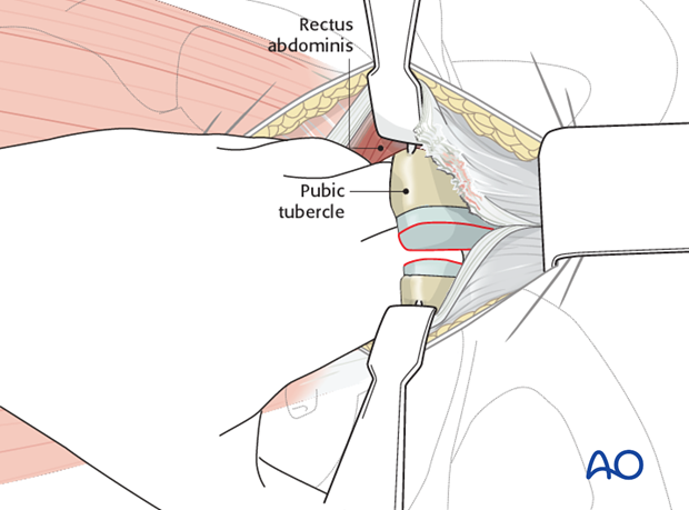
7. Dissect the rectus abdominis
Anteriorly it might be necessary to elevate the recti insertions from the pubic body lateral to identify the obturator foramen.
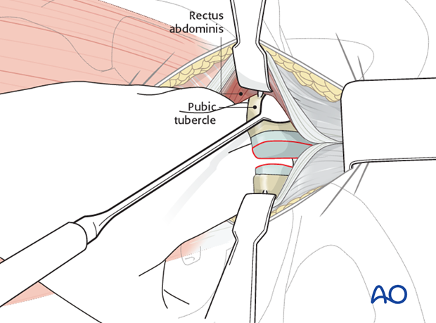
8. Option: insert reduction forceps
If the surgeon plans to place the tips of reduction forceps in or at the edge of the obturator foramina, additional dissection may be carried laterally to expose this area.
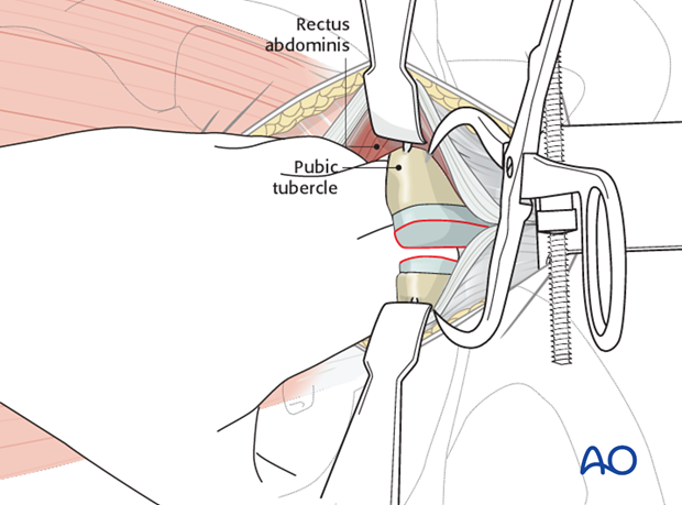
9. Exposure of superior pubic rami
Once the symphysis is exposed, the pubic rami can be identified laterally to the region of the iliopectineal (iliopubic) eminence.
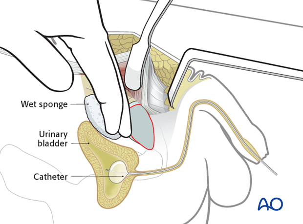
10. Closure
Prior to closure the wound is irrigated and the bladder is inspected for any signs of injury. The urine in the Foley catheter bag is inspected to ensure there is no bleeding.
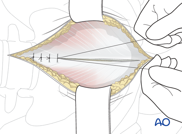
The midline incision in the rectus abdominis and superficial tissues are closed in layers.
