Extraperitoneal pelvic packing
1. Packing versus angio-embolization
In an emergency situation the decision whether to go for packing or angiography with embolization depends on several factors.
- The condition of the patient
- When the patient is in extremis, angiography takes too long
- Availability of direct angiography or an operating room
- Availability of adequate personnel to perform angio-embolization
- When a laparotomy is mandatory, extraperitoneal packing could be part of the same procedure
Combining the two techniques (extraperitoneal pelvic packing and angio-embolization) is an option.
2. Patient positioning
Place the patient in supine position.
A urinary catheter should be inserted prior to beginning the procedure.
In male with a pelvic fracture, urethral ruptures should be excluded before introduction to the catheter to avoid creation of a false route.
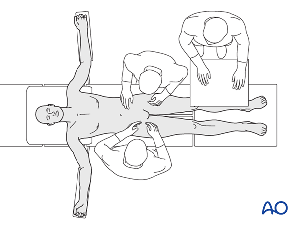
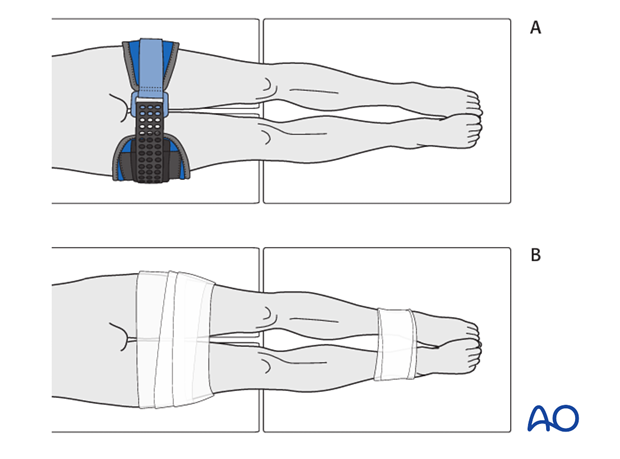
If present, a temporary pelvic binder should be removed and replaced more distally (A).
Internal rotation of the legs can be preserved during the procedure by taping the legs in this position (B) with adequate padding.
For pelvic packing to compress the source of bleeding, the pelvis should be stabilized.
3. Skin incision
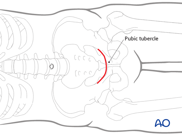
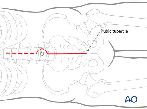
Horizontal incision (“Pfannenstiel”)
A Pfannenstiel incision can be used when there is no obvious bleeding in the abdomen.
Perform a horizontal incision about 2 fingerbreadths proximal to the pubic tubercle.
The length of this incision is typically 5-10 cm. It can be extended further laterally on one or both sides depending on the needed exposure.
Vertical incision
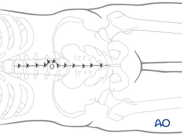
There may be a need for extension of the incision cranially in case of abdominal bleeding. In this case a vertical incision is preferred.
4. Deep dissection
Dissect the subcutaneous tissue and identify the anterior rectus fascia.
Locate the linea alba and incise it longitudinally.
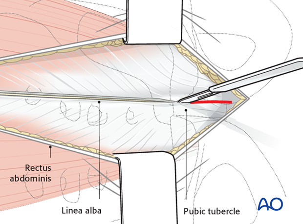
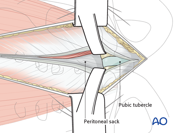
Both bellies of the rectus abdominis muscle are gently retracted laterally.
Identify the peritoneal sack but do not open it.
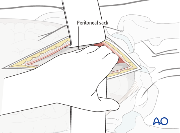
Use your fingers to bluntly retract the peritoneal sack superiorly. It can be dissected as far posteriorly as the SI joints to create retroperitoneal spaces bilaterally. These may already have been torn open by the injury.
Care is taken not to tear the peritoneum.
5. Extraperitoneal pelvic packing
Fill the retroperitoneal spaces with as many big gauzes as possible to create a good pressure on both sides of the retroperitoneal region.
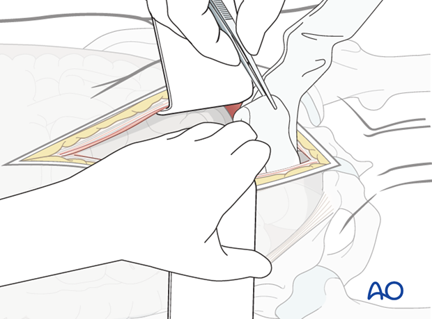
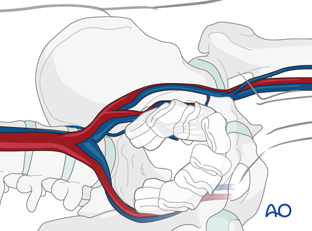
Reduction of the pelvis
To realign and stabilize the pelvis an anterior external fixator or C-clamp is placed.
In case of a disrupted symphysis, immediate reduction and plate fixation can be performed through the same incision when the patient is relatively stable after packing.
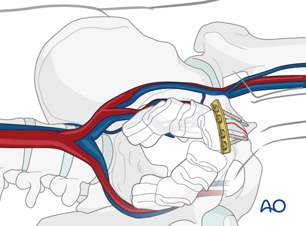
6. Temporary closure
Prior to closure the wound is irrigated and the bladder is inspected for any signs of injury. The urine in the Foley catheter bag is inspected to ensure there is no bleeding.
The fascia of the abdomen is temporarily closed.
Skin incision can be left open and dressed with a large plastic adhesive drape.
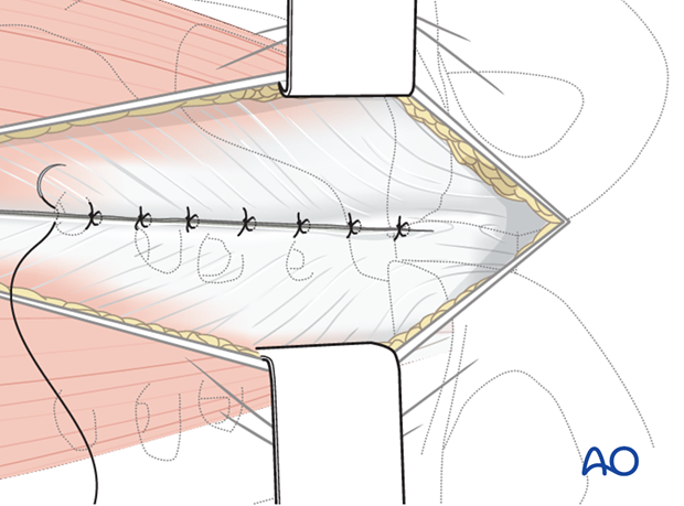
7. Packing removal
A "second look" must be done between 24 and 48 hours. If bleeding has stopped, the packs soaked and gently removed. If bleeding persists, they should be replaced.
8. Final closure
The midline incision in the rectus abdominis is closed in one or two layers. The subcutaneous tissues and skin are then closed in layers.
