External fixation: Emergency stabilization with a C-clamp
1. Preparation of entry point
Orientation and skin incision
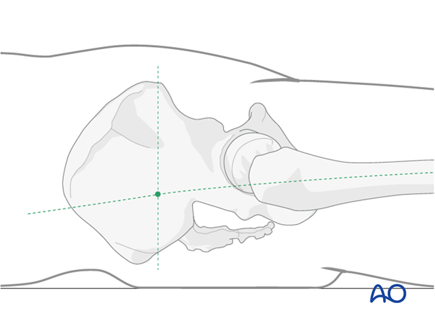
Draw a line from the anterior superior iliac spine to posterior superior iliac spine.
Palpate the greater femoral trochanter and draw a line along the axis of the femoral shaft perpendicular to the first line.
A small skin incision for the entry point is made at the junction of the two lines.
Bone entry point
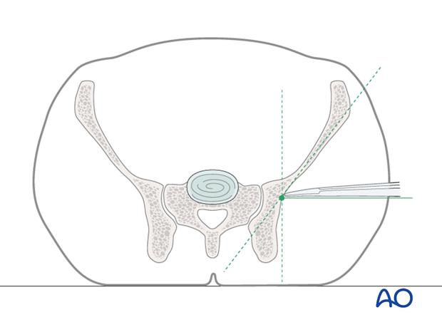
Spread the subcutaneous layers and muscle with scissors till bony contact is reached.
Let the scissors glide into the depression between the oblique orientation of the iliac wing and the vertical part of the posterior iliac wing until the center of the cavity is reached.
This is the ideal entry point directly opposite of the S1 body of the sacrum.
If available, image intensifier should be used in the lateral projection to confirm that the entry point overlies the center of the S1 body.
Teaching video
AO teaching video: Pelvis – Unstable Fractures of the Pelvic Ring – Emergency Stabilization with Pelvic C-clamp
2. Reduction
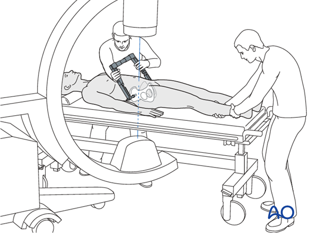
The pelvis should be realigned as well as possible before the C-clamp is applied.
Traction and/or rotation of the leg is used to restore alignment.
Control under image intensification!
3. Clamp positioning
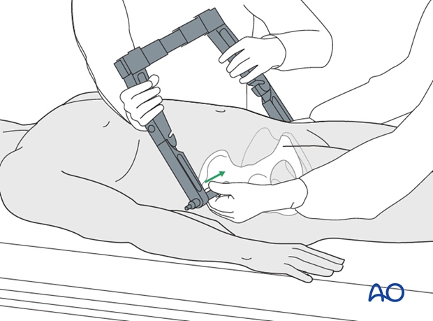
The side struts with attached nails are advanced through the incisions to the bone until the tips have a firm hold in the lateral cortex at the correct entry point.
Compression
With the pelvis realigned and the spikes appropriately positioned, the spike tips are hammered into the outer cortex of the ilium.
The posterior pelvic ring is closed by the C-clamp by manual compression of the side struts in a medial direction.
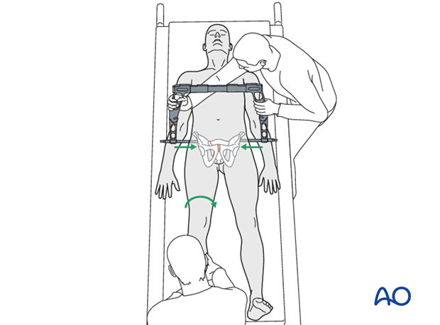
Tightening of screws
The screws are tightened to secure the frame.
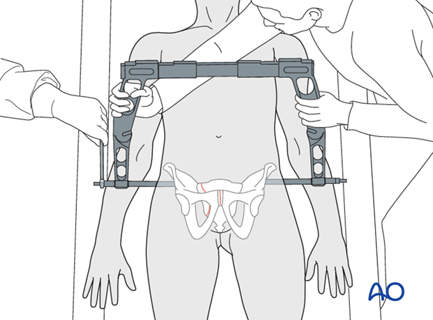
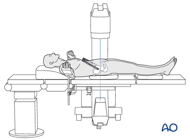
Control reduction and positioning of clamp under image intensification with multiple views.
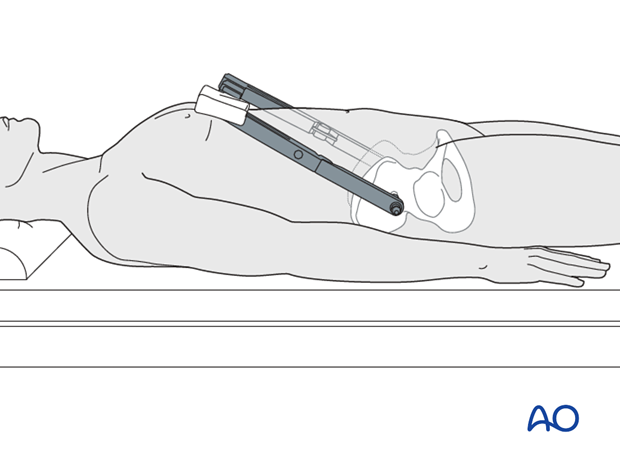
The C-clamp can be turned to make space for laparotomy or diagnostic tools.
Note
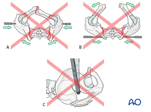
If the C-clamp is positioned too far ventral in the ilium, there may be perforation of this thin part of the ilium or internal rotation of the pelvic ring (A).
If the C-clamp is positioned too far posterior in the ilium, the result will be an external rotation widening of the pelvic ring (B).
Caudal position increases risk of the pins passing through the greater sciatic foramen into the pelvis, injuring nerves and vessels (C).
4. Aftercare following C-clamp fixation
A C-clamp is normally applied as an emergency in patients with pelvic and hemodynamic instability. As soon as the causes of the hemodynamic instability have been addressed, definitive fixation of the pelvis can be performed and the C-clamp removed.


