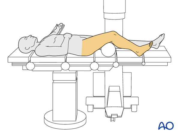
Supine knee flexed 30°
1. Introduction
This position is used for all patellar fractures as the ability to obtain AP and lateral X-rays is exceptional.

2. Preoperative preparation
Operating room personnel (ORP) need to know and confirm:
- Site and side of fracture
- Type of operation planned
- Ensure that operative site has been marked by the surgeon
- Condition of the soft tissues (fracture open or closed)
- Implant to be used
- Patient positioning
- Details of the patient (including a signed consent form and appropriate antibiotic and thromboprophylaxis)
- Comorbidities, including allergies
Antibiotics are administered according to local antibiotic policy and specific patient requirements.
Many surgeons use Gram-positive prophylactic antibiotic cover for closed fractures, adding Gram-negative prophylactic cover for open fractures. Always remember that antibiotic therapy will never compensate for poor surgical technique.
3. Anesthesia
This procedure is performed with the patient under general or regional anesthesia.
If a spinal anesthetic is used, the surgeon and anesthetist need to be confident that the procedure will not last more than 1.5 hours.
Long-lasting postoperative complete pain blocks for the injured leg should be avoided as this could hide symptoms of a subsequent compartment syndrome.
4. Patient and x-ray positioning
- Position the patient supine with roll or well-padded sandbag under the thigh to keep the knee in a 30° flexed position. Alternatively, a carbon triangle may be used.
- Apply a correctly sized tourniquet to the femur with adequate padding.
- Carefully pad all pressure points, especially in the elderly.
- Position the image intensifier on the opposite side of the injury and the surgeon.
- Before preparing and draping, ensure good AP and lateral image intensifier views can be obtained.

5. Skin disinfecting and draping
- Disinfect the exposed area from above the iliac crest to the foot with the appropriate antiseptic.

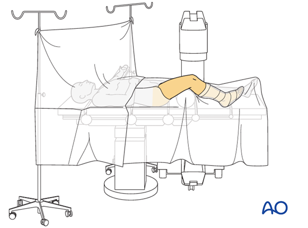
- Drape the limb with a single-use U-drape. A stockinette covers the lower leg and is fixed with a tape. The leg is draped to be freely moved.
- Drape the image intensifier.

6. Operating room set-up
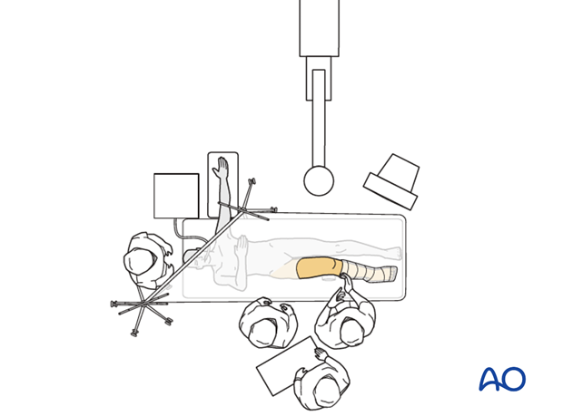
- The surgeon and ORP stand on the side of the affected limb.
- The assistant stands next to the surgeon.
- Place the image intensifier on the opposite side of the injury and the display screen in full view of the surgical team and the radiographer.


