External fixation
1. Principles
Talonavicular joint function
It is crucial to retain TN function as loss of motion:
- Results in loss of complex hindfoot circumduction
- Leads to adjacent joint degeneration (DJD)
Retaining even a small amount of motion is thought to protect the adjacent joint function.
Because of its extensive range of motion, the TN joint is also known as the “coxa pedis.”

Complex fracture
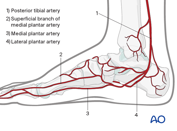
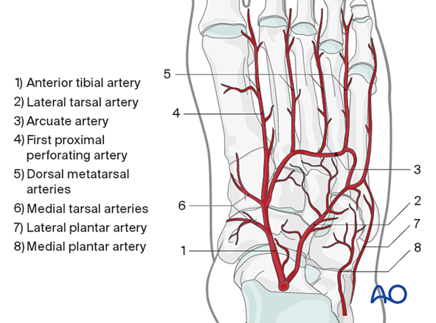
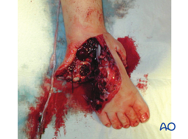
The delicate leash of small vessels around the midfoot is often injured during the original trauma. The vessels and soft tissue must be given time to recover. Soft-tissue defects also need to be addressed.
Maintenance of column length
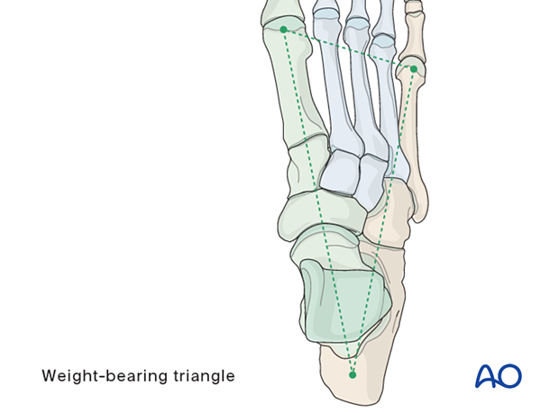
The foot’s form and function depend on the normal relationship between the medial and lateral columns.
Relative shortening of the medial column leads to cavus, whereas relative shortening of the lateral column leads to flat-foot.
If the injury has resulted in comminution with loss of medial or lateral column length, this length and normal geometry must be restored.
Bridging hardware and bone graft will assure proper column length, overall shape, and alignment of the foot.
2. Soft-tissue considerations
External fixation as a temporizing measure
Blisters and soft-tissue injuries are more common in higher-energy injuries and severe injuries to the underlying bone. Usually, surgery is delayed to allow soft tissues to settle before definitive surgical care. Soft tissues must take priority over the bony injury, and once the "wrinkle sign" is present, indicating soft-tissue relative recovery, the incidence of soft-tissue complication from bony surgery is decreased.

External fixation as definitive treatment
Open reconstruction may not be possible in higher-energy injuries and severe injuries to the soft tissue and underlying bone.
In such cases, external fixation with or without percutaneous pins is used to stabilize the foot. This will maintain columnar bony alignment and allow soft-tissue reconstruction. Perhaps at a later date, bony reconstruction may be possible.
The goal of treatment in these injuries is often to maintain a foot that will fit into a shoe and allow ambulation. After severe injuries, the foot may change size and shape. The overall alignment should, however, allow for function.
3. External fixation
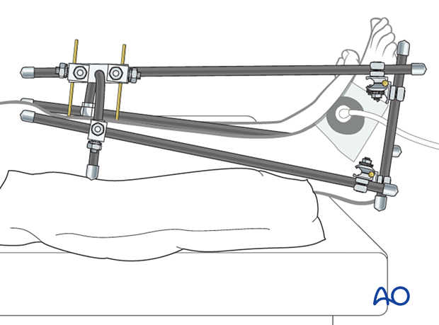
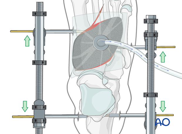
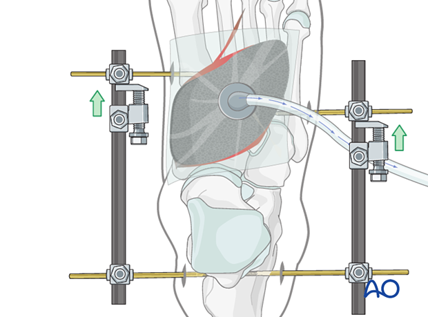
Medial and lateral external fixation (with a distractor device to restore columnar length) should be applied as soon after the injury as possible to stabilize the foot and decrease further injury to the soft tissues.
The soft-tissue injuries can then be addressed.
Antibacterial non-adherent dressings and negative pressure wound therapy (NPWT) can be used as needed.
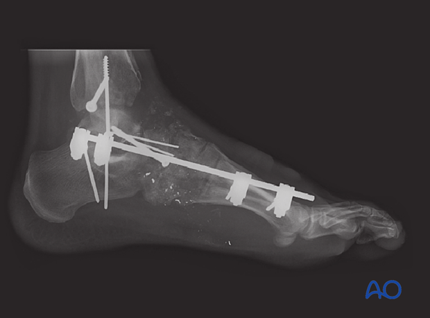
4. Case
AP view of external fixator applied to a midfoot gunshot injury.
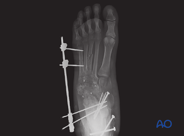
Lateral view of the same case
5. Aftercare
Dressing
A first layer of non-adherent antibacterial dressing is applied in areas not covered by a NWPT dressing.
Hospital protocols are followed for pin care.
Follow-up
The patient should be counseled to keep the leg on a cushion and elevated. The elevation level should be between the waist and the heart to decrease swelling. If the foot is elevated too high (above the heart), it may impede inflow.
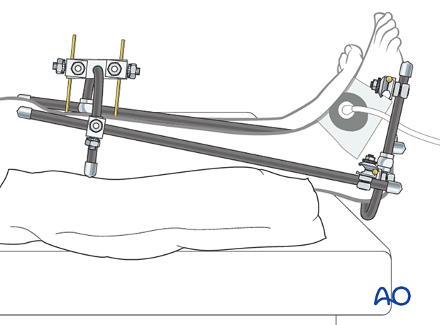
Please note that there are frequently other components to consider in very high-velocity injuries. In this external fixator configuration, the foot's external fixation has been combined with the external fixation of the distal tibia and hind-foot. An anterior bar has been added to prevent equinus deformity.
A gastrocnemius release may need to be performed in cases with postoperative gastrocnemius contracture. This occurs more typically in the mid and hind-foot.
