Intertarsal crush injury
Introduction
Intertarsal injuries are limited by the Lisfranc region distally and the Chopart region proximally. It may involve the navicular-cuneiform or intercuneiform joints of the midfoot or the cuneiform bones.
History and physical exam
These injuries may arise in athletics or result from higher-energy injuries. Their mechanism is similar to a Lisfranc midfoot injury, or they may indeed be part of Lisfranc fracture-dislocation.
The high-energy injury is often part of a polytrauma and is often associated with other injuries in the foot and other body parts.
In the multiply-injured patient, intertarsal injuries and fractures may be picked up in the secondary survey. In the case of unconscious patients, care should be taken to check for unusual swelling, crepitus and/or deformity. If suspected, foot x-rays are indicated.
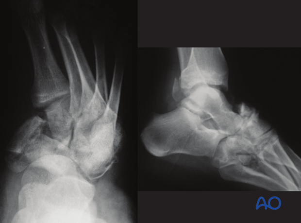
This x-ray shows a severe injury of the foot with complete disorganization of the anatomy.

High-energy injuries are often associated with other injuries, both in the foot and the rest of the body.
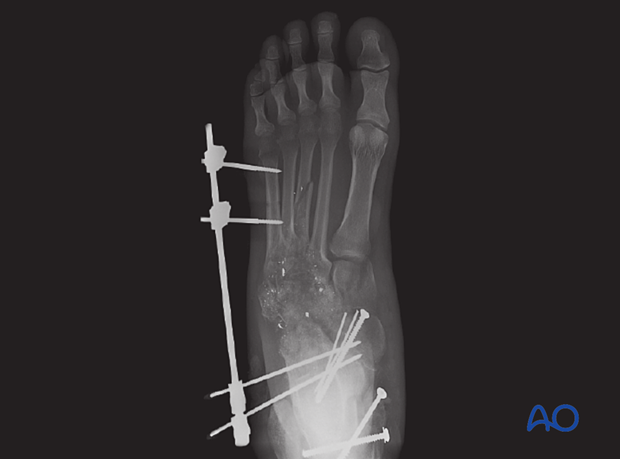
In the event of severe soft-tissue injury, possibly with vascular compromise, or comorbidities (diabetes, etc.), external fixation and/or percutaneous pinning may be the only treatment choice available, at least in the short term. Much later, reconstruction may be possible.
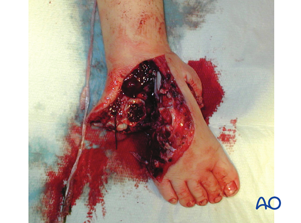
This clinical photograph shows a severe open fracture.

Imaging
Plain x-rays will often show the injury pattern.
CT with sagittal and coronal reconstruction is useful in obtaining a three-dimensional understanding of the injury. CT protocol should be thin cuts with large overlap.


