Patient assessment
1. Clinical findings
Clinical examination should evaluate:
- Swelling
- Deformities
- Stability
- Plantar ecchymosis
- Location of pain
Swelling
What is observed:
- General swelling of the foot
What it indicates:
- Potential compartment syndrome
- Any bony or ligamentous midfoot injury
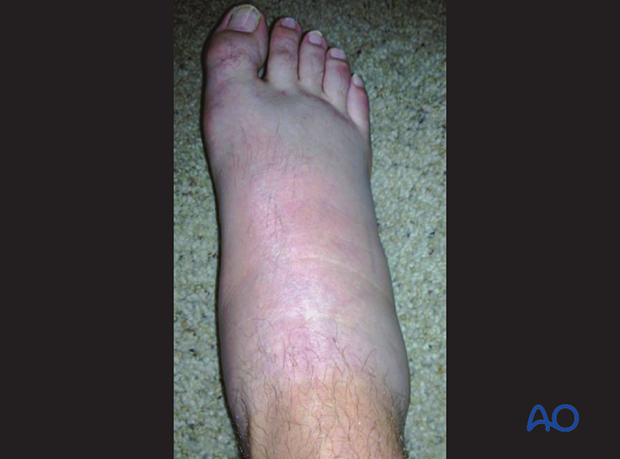
Good case of normal limb on left and swelling on the right foot
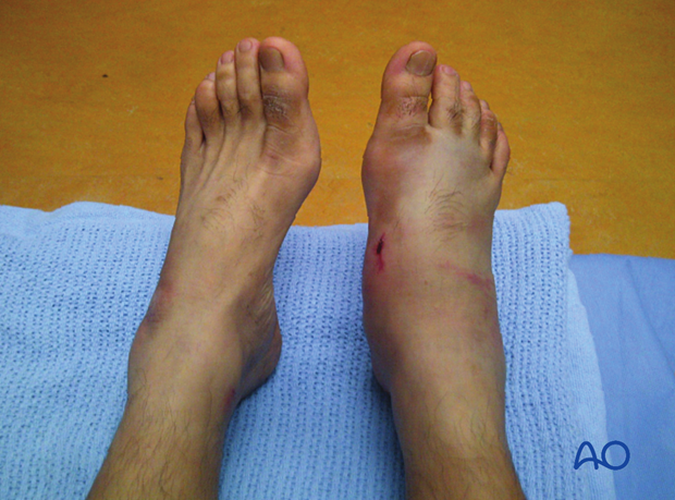
Deformities
Varus/valgus midfoot deformityWhat is observed:
- Varus midfoot deformity
What it indicates:
- Cuneiform, navicular, Lisfranc and/or Chopart injury
What is observed:
- Valgus midfoot deformity
What it indicates:
- Cuboid, Lisfranc and/or Chopart injury
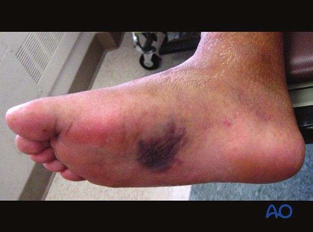
What is observed:
- Plantar ecchymosis sign
What it indicates:
- Any bony or ligamentous midfoot injury
Instability testing
Instability testing will most often need to be performed with a regional block or under general anesthesia.
Instability dictates injuries to bone and/or ligaments.
Pain location
Pain will indicate injuries to underlying structures.
2. Radiographs
The following views may be taken:
- Lateral view
- AP view
- 45° oblique view
- Stress view
- Weight-bearing view
Lateral view
What is observed:
- Subtle dorsal displacement of a metatarsal base
What it indicates:
- Ligament injury to the Lisfranc joint
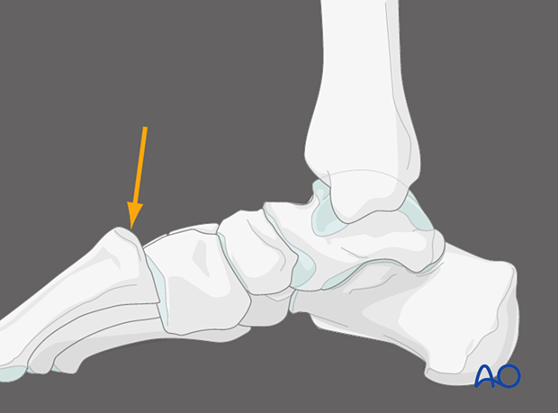
This lateral x-ray shows a subtle dorsal positioning of a metatarsal base.
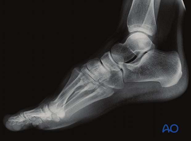
What is observed:
- Step at Lisfranc and Chopart joint levels
What it indicates:
- Lisfranc and Chopart injury
This lateral x-ray shows a significant injury through the midfoot involving the Chopart and Lisfranc joints
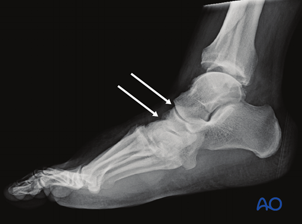
45° Oblique
This is the best view to identify cuboid fractures.
What is observed:
- Malalignment at the medial border of cuboid and 4th metatarsal
What it indicates:
- Lisfranc injury
- Disrupted lateral column
- Metatarsal fractures
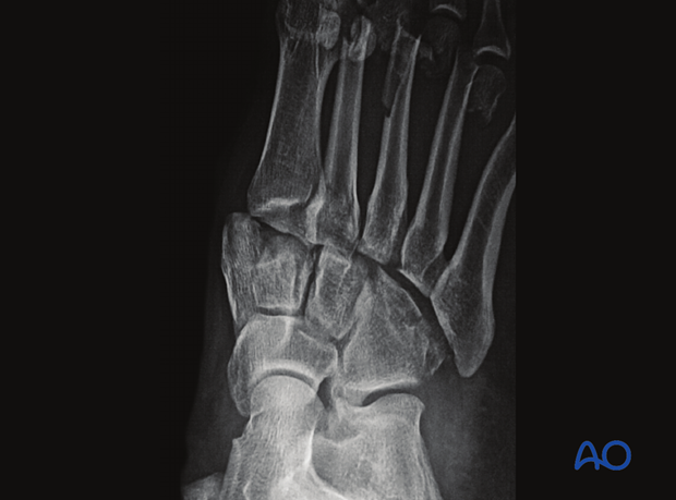
AP view
What is observed:
- Malalignment at the medial border of intermediate cuneiform and the 2nd metatarsal
What it indicates:
- Lisfranc injury
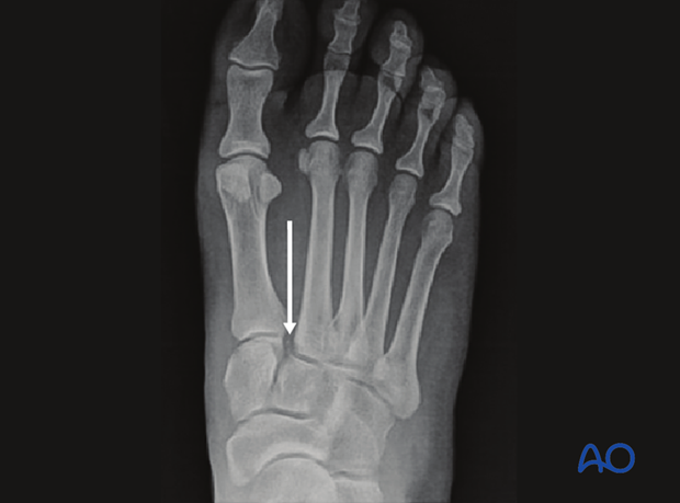
Another example of the same radiological pattern
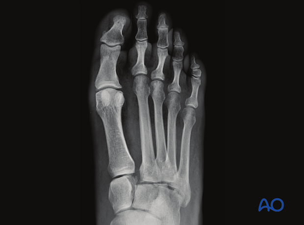
What is observed:
- Fracture at the base of 2nd metatarsal
What it indicates:
- Lisfranc injury
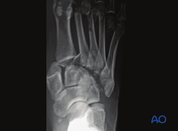
What is observed:
- >3 mm distance between the base of 2nd and 3rd metatarsal
What it indicates:
- Lisfranc injury
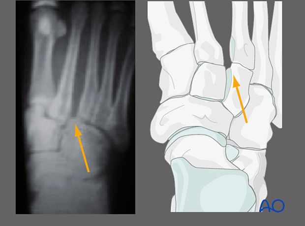
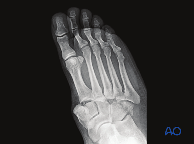
What is observed:
- Tarsometatarsal dissociation
What it indicates:
- Lisfranc injury
Stress views
Stress views can be obtained using an image intensifier either using a regional block or intraoperatively under anesthesia.
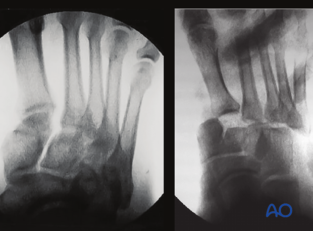
Weight-bearing views
Weight-bearing views allow better visualization of dissociation and instability in the lateral, AP, and 45° oblique views.
This is particularly useful in pure ligamentous injuries.
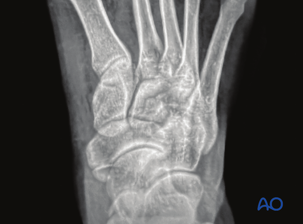
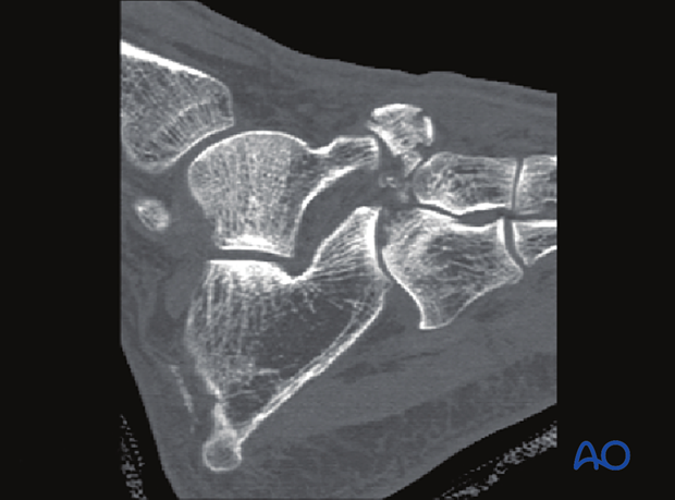
3. CT imaging
What is seen:
- Small avulsion fracture of the navicular from the “constant fragment”
What it indicates:
- Possible instability of the Chopart joint
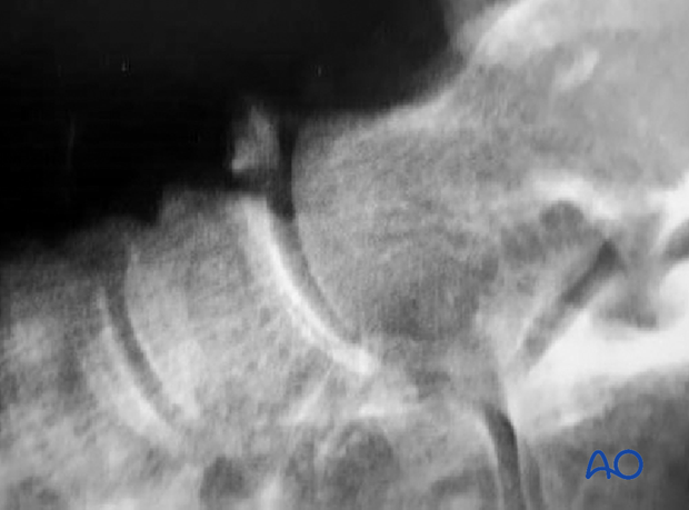
What is seen:
- Fracture dislocation of the Chopart joint
What it indicates:
- Unstable Chopart joint
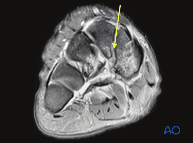
4. MRI
MRI in the acute situation is only useful when no osseous injuries are found.
What is seen:
- Ruptured Lisfranc ligament
- Dissociation of the navicular from the “constant fragment”
What it indicates:
- Lisfranc injury
